
|
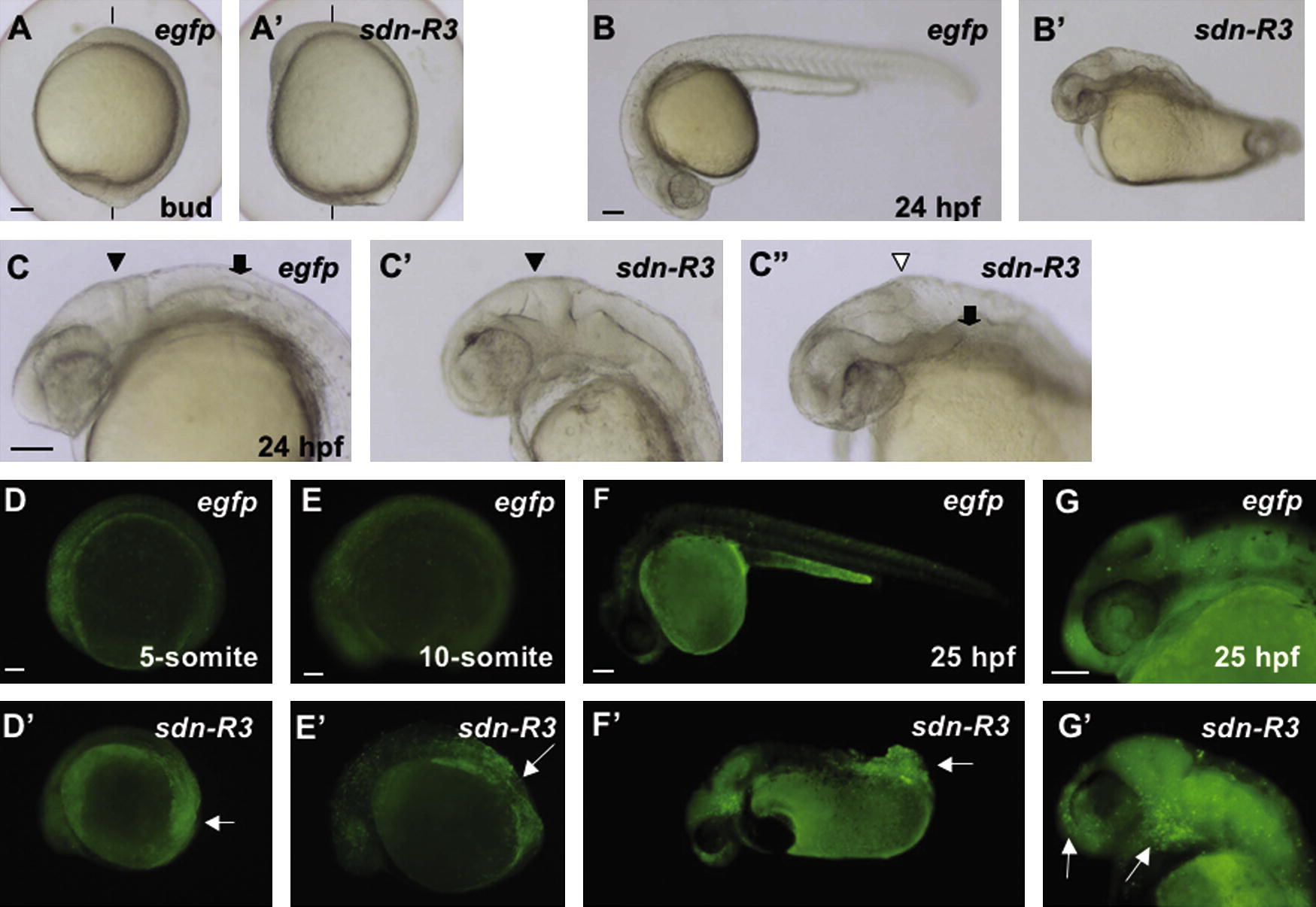
Fig. 8 Effects of sdn-R3 overexpression on zebrafish embryos. (A–C, A′–C′, and C″) Bright-field views of embryos injected with mRNA for egfp (A–C, 300 pg/embryo) or sdn-R3 mRNA (C′), 100 pg/embryo; A′, B′, and C″, 300 pg/embryo). (A and A′) Animal–vegetal axes are shown with vertical lines. (D–G and D′–G′) Epifluorescence views of embryos injected with mRNA for egfp (D–G) or sdn-R3 (D′–G′) (300 pg/embryo). Normal isthmic constrictions, absence of the isthmus, otic vesicles, and accumulation of apoptotic cells are marked with sold triangles, open triangles, thick arrows, and thin arrows, respectively. Lateral views with anterior to the top and dorsal to the right (A and A′) or with anterior to the left and dorsal to the top (all the other panels) are shown. Scale bars, 100 μm.
Reprinted from Mechanisms of Development, 126(1-2), Ota, S., Tonou-Fujimori, N., and Yamasu, K., The roles of the FGF signal in zebrafish embryos analyzed using constitutive activation and dominant-negative suppression of different FGF receptors, 1-17, Copyright (2009) with permission from Elsevier. Full text @ Mech. Dev.