
|
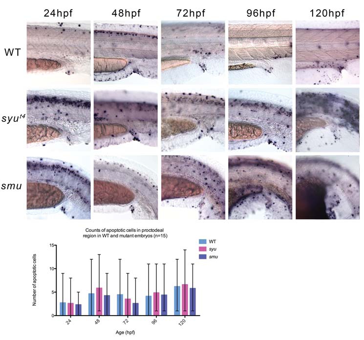
Fig. 6 Apoptotic events in the developing posterior gut of wild type and Hh pathway mutants. (A-E) TUNEL staining of WT embryos between 24-120 hpf reveals the normal pattern of apoptosis during the formation of the posterior gut and pronephric ducts. At 120 hpf a number of cells in the cloaca die creating an externally opening posterior gut. (F-J) TUNEL staining of syu mutant between 24-120 hpf. (K-O) TUNEL staining of smu mutants between 24-120 hpf. (P) Counts of apoptotic cells in the mutants and WT siblings reveals that there are large differences in the amount of cell death between embryos of the same age. However, there is no significant difference between the number of apoptotic cells in WT siblings and mutant embryos.