
|
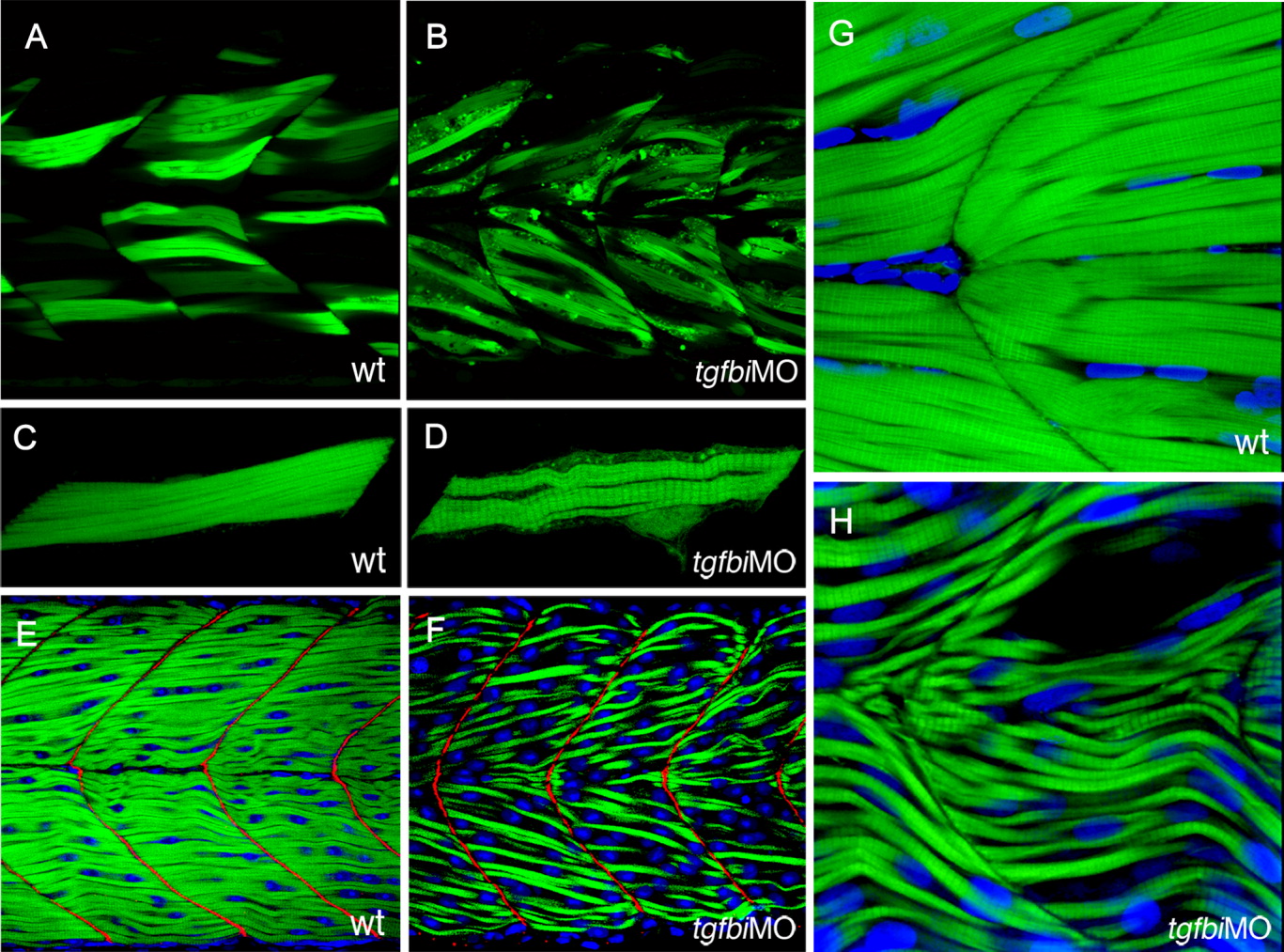
Fig. 3 Attachment of muscle fibres to the myoseptum is not disrupted by Tgfbi knock-down. A-D: Muscle fibres in wild type and tgfbi morphant embryos that have been scatter labelled by transient expression of the α-actin:GFP transgene and imaged at 50 hpf. Like their wild-type counterparts (A), the labelled myofibres in tgfbi morphants (B) spanned entire somites with no disconnected muscle fibres. Projections of 3D images of individual labelled fibres show that the morphant muscle fibres (D) were thinner and wavier than their wild type counterparts (C). E-H: Embryos that have been stained with FITC-conjugated Phalloidin (green) to label muscle fibres and Topro3 (blue) to label nuclei. In E and F, staining for Vinculin (red) reveals that the vertical myosepta were not affected by the loss of Tgfbi. The fibres in morphant embryos (H) were much thinner and less closely packed than in wild type (G) with some empty spaces between them.