
|
Fig. S4
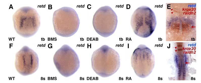
RA Signaling Positively Regulates retd in the LPM
Expression of retd at the tailbud (tb) or 8 somite stages, dorsal views, anterior to the top. (A-D and F-I) Reduction of RA signaling inhibits retd expression, and treatment with RA induces ectopic retd expression. (E) At tb, retd expression (blue) overlaps with raldh2 expression (red-brown; arrow). (J) By the 8 somite stage, retd expression is anterior to the expression of raldh2 in the somites (arrow). krox20 expression provides a marker of hindbrain rhombomeres 3 and 5 (anterior brown stripes; arrowheads). RA treatment is known to positively regulate retd expression in cell culture (Cerignoli et al., 2002), but it has not been previously shown that RA signaling is required for its expression in the embryo nor has its expression been previously reported in zebrafish. Based on its proximity to raldh2 and three retinoic acid response elements in the 2 kb 5′ to the retd start site (J.S.W. and D.Y., unpublished data; Bastien and Rochette-Egly, 2004), it is feasible that retd is a direct target of RA signaling.
Reprinted from Developmental Cell, 15(6), Waxman, J.S., Keegan, B.R., Roberts, R.W., Poss, K.D., and Yelon, D., Hoxb5b acts downstream of retinoic Acid signaling in the forelimb field to restrict heart field potential in zebrafish, 923-934, Copyright (2008) with permission from Elsevier. Full text @ Dev. Cell