
Image
|
Figure Caption
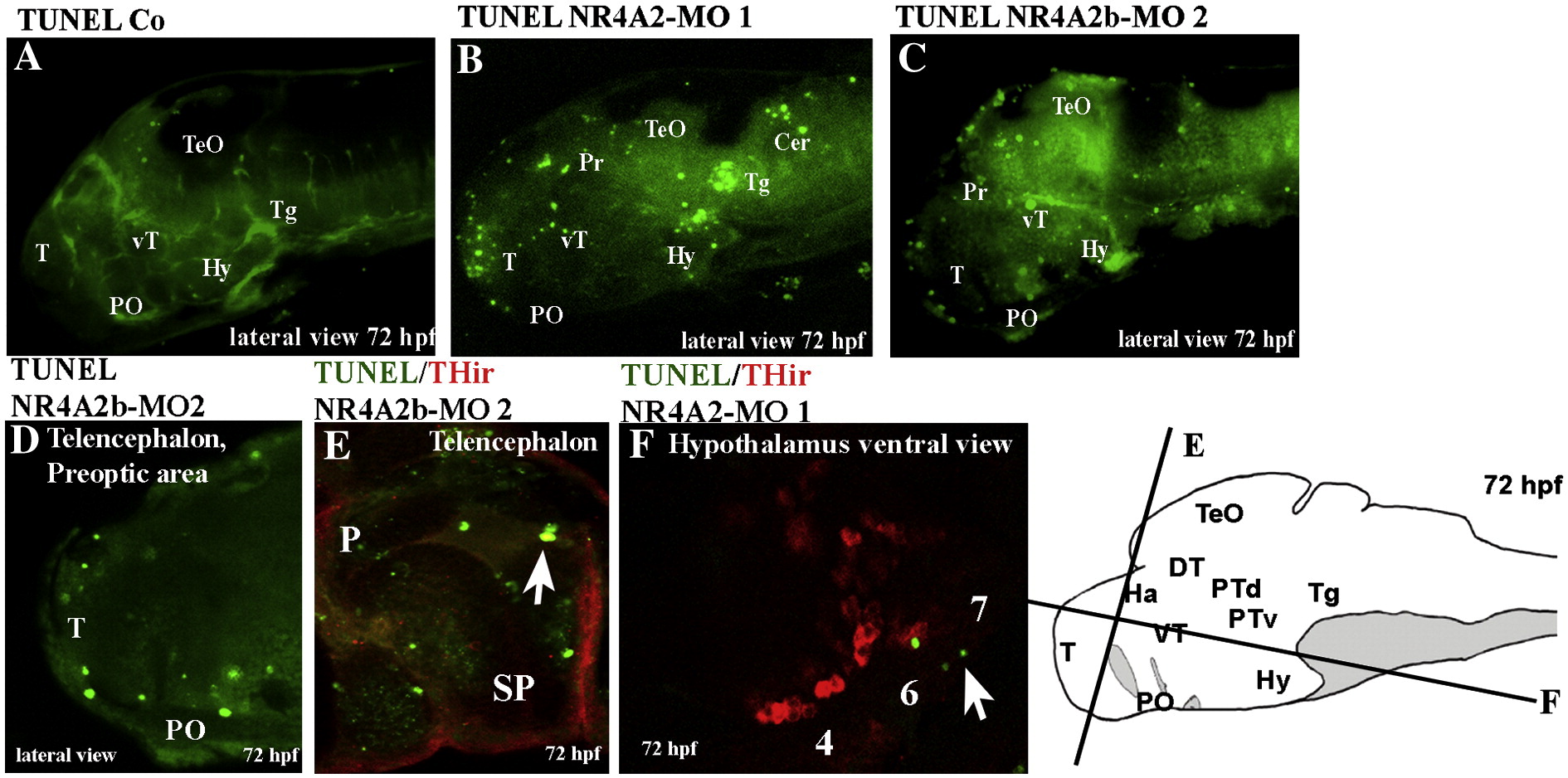
Fig. 7 Apoptosis in TH-expressing cells in controls and morphant larvae at 72 hpf. In all panels, apoptotic cells are stained in green following TUNEL detection on control larvae (A), NR4A2-MO1-injected larvae (B,F) and NR4A2b-MO2-injected larvae (C–E). Panels E and F depict in addition a double staining for TH protein (immunocytochemistry, red). A–D are whole-mount views, anterior to the left, dorsal up; E,F are sections (planes indicated on the schematic, right panel) photographed under confocal microscopy.
Figure Data
Acknowledgments
This image is the copyrighted work of the attributed author or publisher, and
ZFIN has permission only to display this image to its users.
Additional permissions should be obtained from the applicable author or publisher of the image.
Reprinted from Molecular and cellular neurosciences, 39(4), Blin, M., Norton, W., Bally-Cuif, L., and Vernier, P., NR4A2 controls the differentiation of selective dopaminergic nuclei in the zebrafish brain, 592-604, Copyright (2008) with permission from Elsevier. Full text @ Mol. Cell Neurosci.