
Image
|
Figure Caption
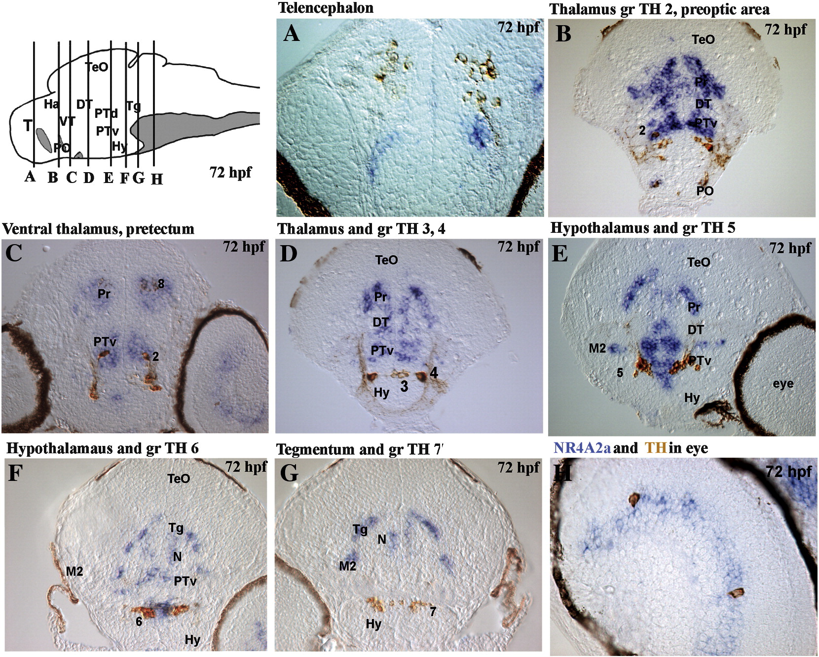
Fig. SD12 Compared expression of NR4A2a and TH during brain development. Localization of NR4A2a transcripts obtained by in situ hybridization (blue staining) and of TH protein obtained by immunochemistry (brown). All views are cross-sections of 72 hpf larvae, dorsal up, in order along the antero-posterior axis (except J: eye). The brain drawing (top left panel) shows the position of the sections. The numbers refer to TH cell groups (Rink and Wullimann, 2002).
Acknowledgments
This image is the copyrighted work of the attributed author or publisher, and
ZFIN has permission only to display this image to its users.
Additional permissions should be obtained from the applicable author or publisher of the image.
Reprinted from Molecular and cellular neurosciences, 39(4), Blin, M., Norton, W., Bally-Cuif, L., and Vernier, P., NR4A2 controls the differentiation of selective dopaminergic nuclei in the zebrafish brain, 592-604, Copyright (2008) with permission from Elsevier. Full text @ Mol. Cell Neurosci.