
Image
|
Figure Caption
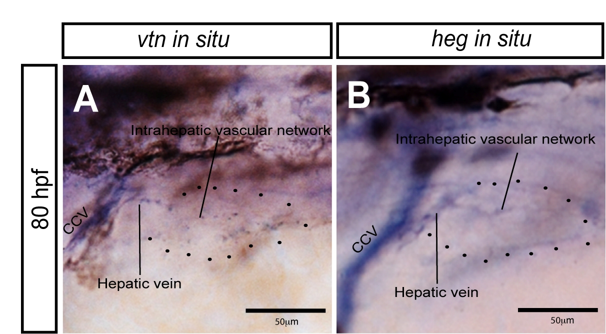
Fig. S7 vtn and heg expression in the liver. (A, B) Higher magnification views of vtn (A) and heg (B) expression in 80 hpf wild-type larvae. Lateral views, anterior to the left. Black dots outline the position of the liver. Both genes are expressed in the common cardinal vein and this expression pattern is continuous with the hepatic vein and intrahepatic vascular network. Thus, both genes appear to be expressed in endothelial cells in the liver. CCV, common cardinal vein.
Acknowledgments
This image is the copyrighted work of the attributed author or publisher, and
ZFIN has permission only to display this image to its users.
Additional permissions should be obtained from the applicable author or publisher of the image.
Full text @ Curr. Biol.