
|
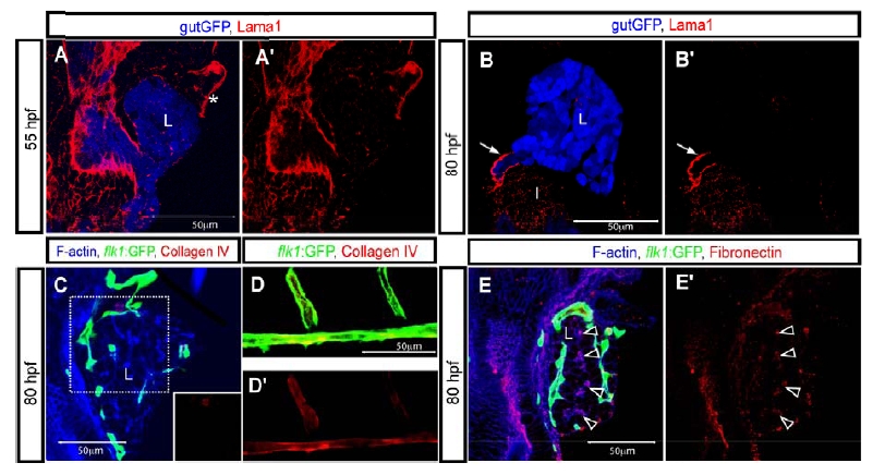
Fig. S5 Extracellular matrix protein deposition in the developing liver. (A and B) z-plane confocal images of a Tg(gutGFP)s854 larva visualized for GFP (pseudo-colored blue) and Lama1 (Laminin alpha 1; red) expression at 55 (A) and 80 (B) hpf. Lama1 immunostaining is shown separately in A′ and B′. Endothelial cells contacting the anterior side of the liver appeared to be surrounded by Lama1 expression (asterisk) at 55 hpf. Lama1 is strongly deposited around the extrahepatic biliary duct (arrow), but not significantly inside the liver at 80 hpf. However, it is worth noting that Lama1 is not the major laminin isoform expressed by endothelial cells26. (C) z-plane confocal image of Tg(flk1:EGFP)s843 liver visualized for GFP (green), F-actin (blue) and Collagen IV (red) at 80 hpf. Collagen IV immunostaining in the outlined area is shown separately in bottom right corner. No Collagen IV immunostaining is detected in the liver. (D) z-plane confocal image of the dorsal aorta and intersomitic vessels of a Tg(flk1:EGFP)s843 larva. The larva was visualized for GFP (green) and Collagen IV (red) expression at 80 hpf. Collagen IV immunostaining is shown separately in D′. Collagen IV surrounds the dorsal aorta and intersomitic vessels. (E) z-plane confocal image of a Tg(flk1:EGFP)s843 larva visualized for GFP (green), F-actin (blue) and Fibronectin (red) at 80 hpf. Fibronectin immunostaining is shown separately in E′. Fibronectin deposition is detected in, or around, the intrahepatic biliary network (black arrowheads). L, liver; I, intestine.