
Image
|
Figure Caption
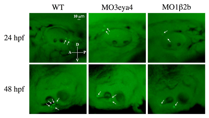
Fig. S2 Acridine Orange staining of otic vesicles from 24 and 48 hpf wild-type, eya4 and atp1b2b morphant fish. Positive signals (indicated by white arrows) were observed in a small number of embryos. Those embryos with maximal staining from each group at both time points are presented. In general, only background levels of Acridine Orange staining were observed in wild type, and eya4 and atp1b2b morphants.
Acknowledgments
This image is the copyrighted work of the attributed author or publisher, and
ZFIN has permission only to display this image to its users.
Additional permissions should be obtained from the applicable author or publisher of the image.
Full text @ Development