
|
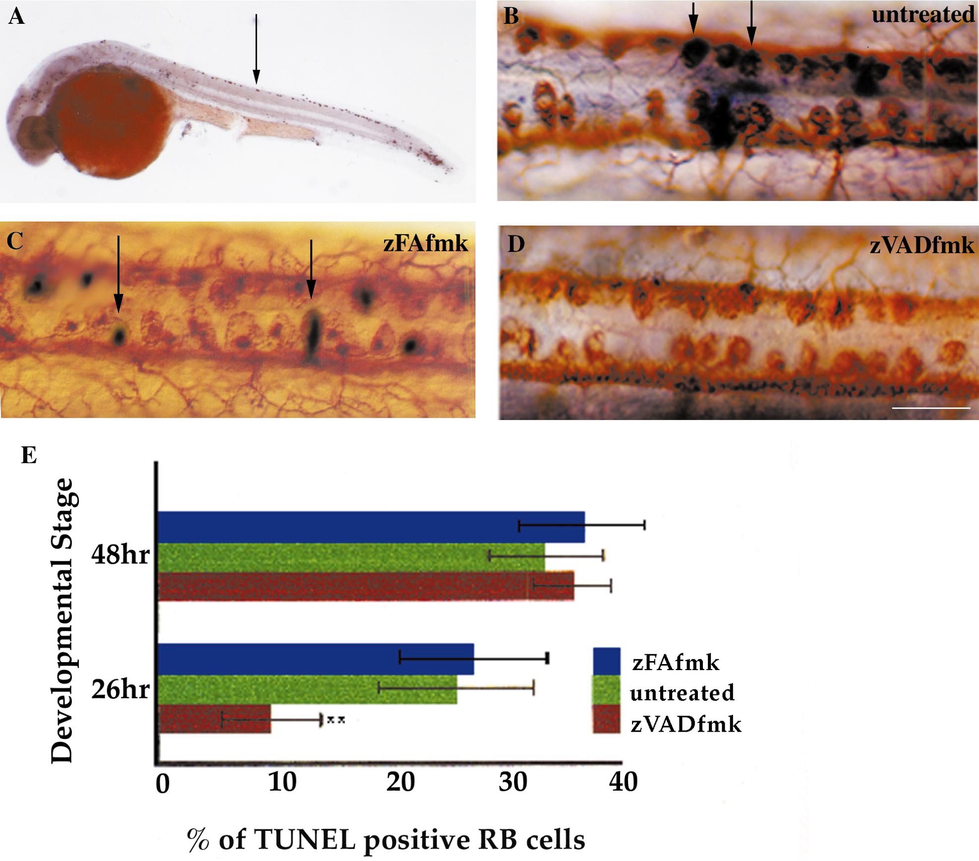
Fig. 2 Rohon Beard neurons die by caspase-dependent programmed cell death. (A–D) Dorsal views of spinal cords at 26 hpf (B–D) or a lateral view of a whole embryo at 28 hpf (A). (A) TUNEL labeling reveals a row of large apoptotic cells (arrow) within the dorsal spinal cord. (B) Double labeling with HNK-1 antibody (brown) and TUNEL (black) shows that a subpopulation of RB cells is dying. Two double-labeled cells are indicated with arrows. (C) zFAfmk (a control peptide)-treated embryo double labeled with HNK-1 (brown) and TUNEL (black). Double-labeled cells (arrows) are found in a frequency similar to that of wild-type embryos. (D) Caspase inhibitor zVADfmk-treated embryo double labeled with HNK-1 (brown) and TUNEL (black). There are no dying cells in this example. Scale bar, 25 μm. (E) Graph showing that the caspase inhibitor zVADfmk significantly reduces programmed cell death of RB cells in 26-hpf but not 48-hpf embryos compared to untreated and zFAfmk-treated controls. Values represent means ± SEM of five separate determinations. **P < 0.02, according to the Student t test.
Reprinted from Developmental Biology, 226(2), Williams, J.A., Barrios, A., Gatchalian, C., Rubin, L., Wilson, S.W., and Holder, N., Programmed cell death in zebrafish Rohon Beard neurons is influenced by TrkC1/NT-3 signaling, 220-230, Copyright (2000) with permission from Elsevier. Full text @ Dev. Biol.