
|
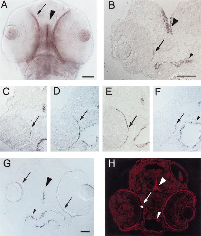
Fig. 1 Expression of shh and twhh in the RPE of embryonic zebrafish. (A) PTU-treated, 56-hpf embryo, ventral view of whole mount. shh is expressed at the ventral midline of the developing CNS (arrowhead), in the eye (arrow), and also in the endothelial lining of the foregut (not in focus in the photo but visible as a purple haze below/between the eyes). Scale bar (applies to A, C, D, E, F, and H), 50 μm. (B) Frontal cryosection of a PTU-treated 45-hpf embryo, hybridized with shh cRNA. Expression is evident in the CNS (large arrowhead), in the foregut (small arrowhead), and in a highly restricted pattern of expression in the RPE (arrow). Nearby (posterior) sections passed through the optic nerve (not shown). Scale bar, 50 μm. (C–F) Cryosections of PTU-treated, 54-hpf embryos hybridized with shh (C, E, and F) or twhh (D) cRNA. C, D, and F are sections from the same embryo, C is the most anterior, F is the most posterior. E is a section from a different embryo, but with sectioning depth and orientation corresponding to a region in between D and F. In both embryos, the CNS midline (large arrowheads) and foregut (small arrowheads) are labeled, but the extent to which the signal extends across the RPE varies with the anterior–posterior level of the section. (G) Oblique, frontal cryosection of a PTU-treated 81-hpf embryo, hybridized with shh cRNA; section cuts through the middle of the left eye (right side of photo), but only grazes the ventral surface of the right eye. shh expression is evident in the CNS (large arrowhead), the RPE (arrows), and the foregut (small arrowhead). Scale bar, 50 μm. (H) Cryosection of a PTU-treated, 54-hpf embryo, labeled with a polyclonal rabbit anti-(rat) Hh antibody using indirect cyanine (Cy3) immunofluorescence; large arrowhead indicates labeling of the CNS midline, small arrowhead depicts labeling of the foregut, arrow indicates labeling of the RPE. Some Hh immunoreactivity is present on apical surfaces of developing photoreceptors that are separated from the RPE by the subretinal space (*).
Reprinted from Developmental Biology, 220(2), Stenkamp, D.L., Frey, R.A., Prabhudesai, S.N., and Raymond, P.A., Function for hedgehog genes in zebrafish retinal development, 238-252, Copyright (2000) with permission from Elsevier. Full text @ Dev. Biol.