
|
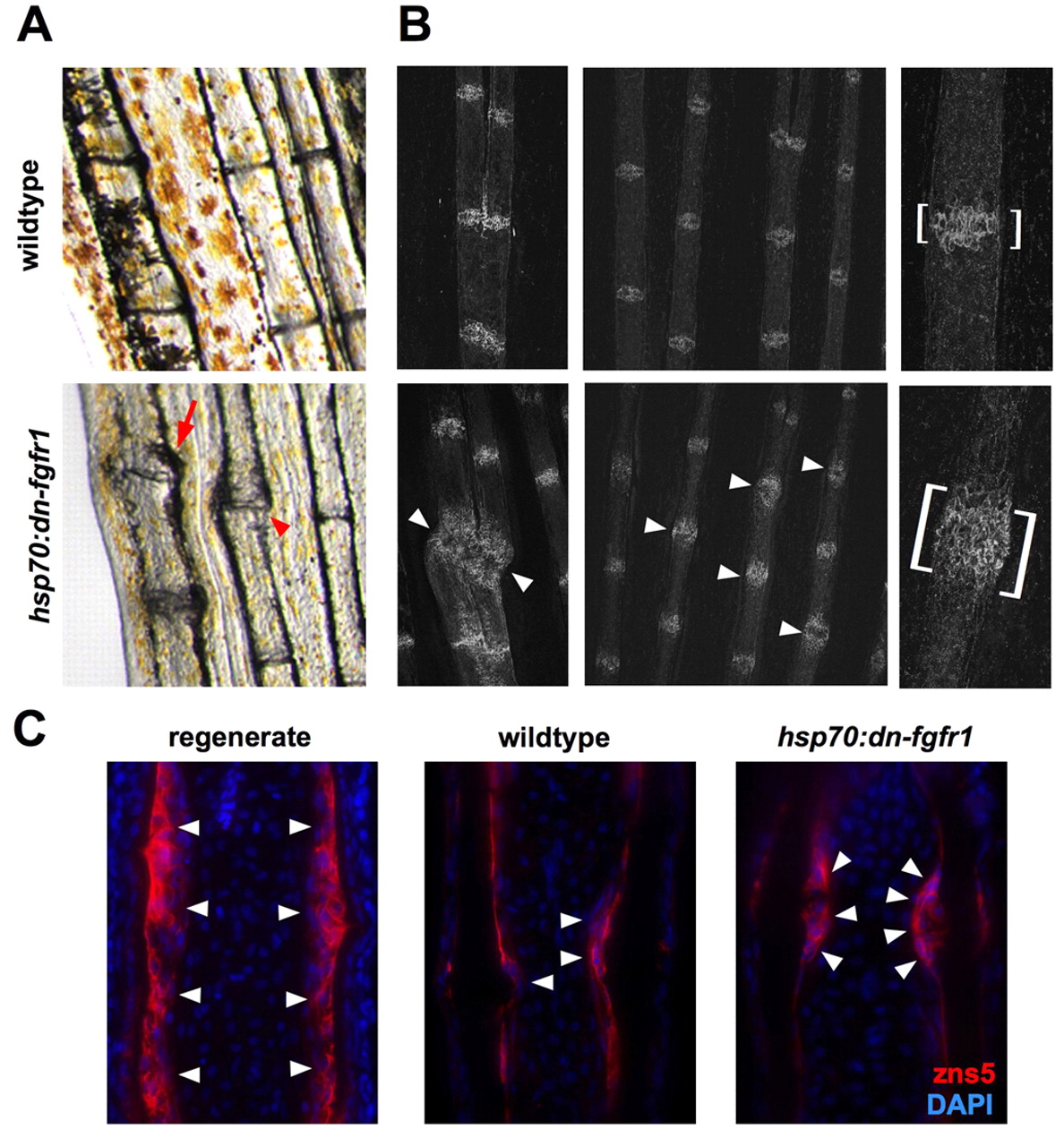
Fig. 2 Fgf receptor inhibition causes pathology at intersegmental joints. (A) Many hsp70:dn-fgfr1 fins exhibited swelling (arrow) or dislocation (arrowhead) of the ray segments at the intersegmental joints (bottom). A representative image of a wild-type fin is provided for comparison (top). (B) Confocal images of whole-mount hsp70:dn-fgfr1 and wild-type fins after 30 days of heatshock, stained with zn3 antibody to visualize scleroblasts. (Left) Example of scleroblast expansion at hsp70:dn-fgfr1 segment joints (bottom) in a case of visible joint pathology (arrowheads). (Middle) Joint hypertrophy was also observed in regions of hsp70:dn-fgfr1 fins without obvious structural damage (arrowheads). (Right) Segmental joints viewed at high magnification, with expansion and disorganization in an hsp70:dn-fgfr1 joint (brackets). (C) Sections of hsp70:dn-fgfr1 and wild-type fins after 30 days of heatshock, stained to visualize scleroblasts with zns5 antibody. There is an expanded zone of rounded scleroblasts (red) surrounding the segment joints of transgenic hemirays. (Left) Regenerates at 4 days post-amputation also have rounded scleroblast morphology.