
|
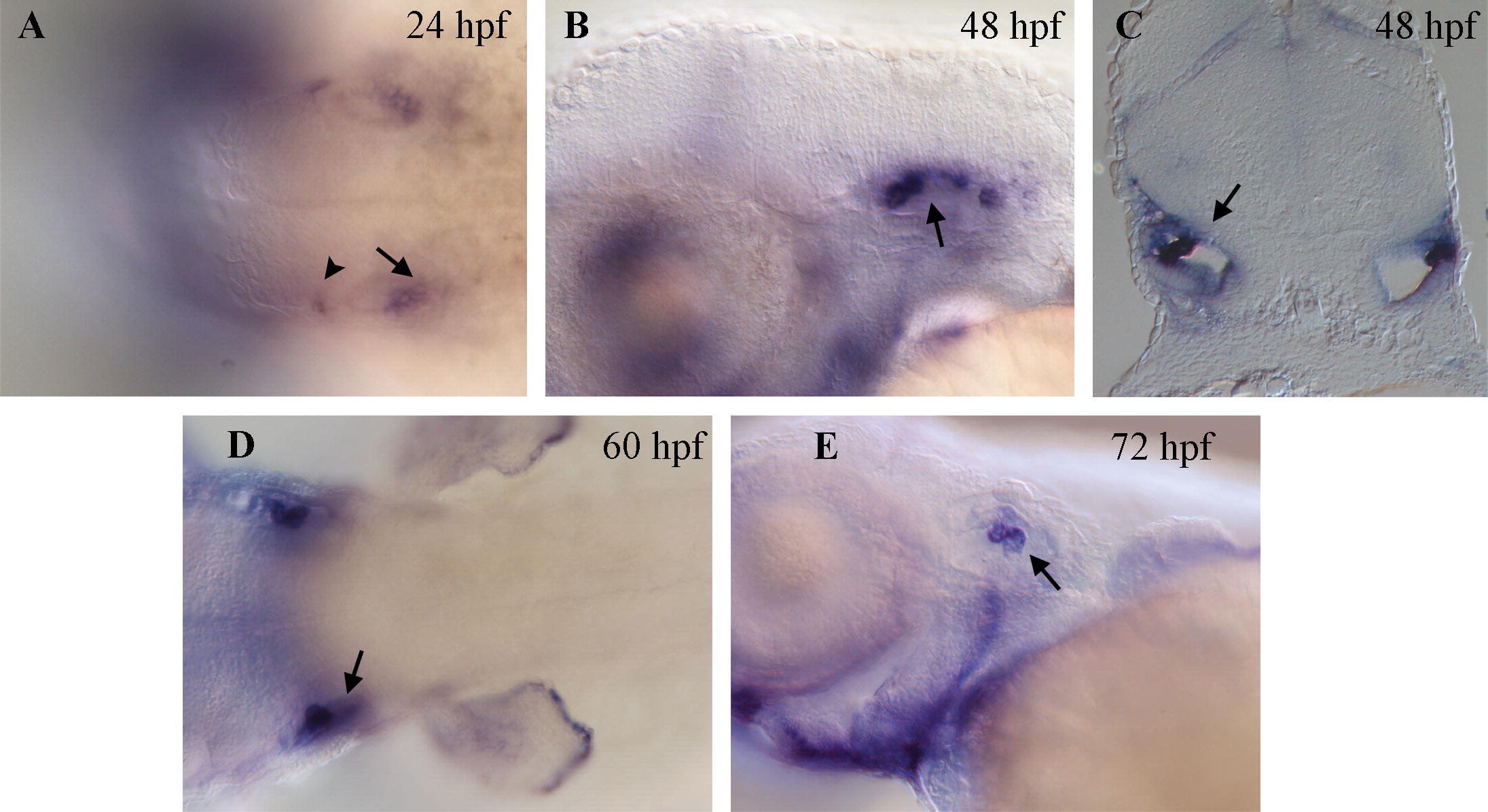
Fig. 4 Expression of bmp7b detected by in situ hybridization in the developing ear. (A) Transcripts of bmp7b can be seen at 24 hpf in the posterior half of the otic vesicle (arrow) and at its anterior most point (arrowhead). (B) Lateral view and (C) cross sectional view of the developing ear at 48 hpf. Strong bmp7b expression can be seen in the forming semi-circular canals (arrows) and weaker expression is observed throughout the epithelium. Expression remains strong in the semi-circular canals at 60 hpf (D, arrow) and 72 hpf (E, arrow) when little or no expression remains in the epithelial component. Anterior is to the left in all panels showing whole mount embryos.
Reprinted from Gene expression patterns : GEP, 8(6), Shawi, M., and Serluca, F.C., Identification of a BMP7 homolog in zebrafish expressed in developing organ systems, 369-375, Copyright (2008) with permission from Elsevier. Full text @ Gene Expr. Patterns