
Image
|
Figure Caption
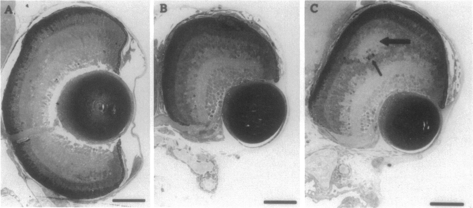
Fig. 5 Absence of a ventral retina as shown by transverse sections through the eyes of 5-day control embryo (A) and embryos which had been treated with 250 μM citral for 2 hr starting at the 2- to 4-somite stage (B and C). Large arrow points to secondary inner plexiform layer. Small arrow points to degenerating photoreceptor cells. (Bars = 50 μm.)
Acknowledgments
This image is the copyrighted work of the attributed author or publisher, and
ZFIN has permission only to display this image to its users.
Additional permissions should be obtained from the applicable author or publisher of the image.
Full text @ Proc. Natl. Acad. Sci. USA