
Image
|
Figure Caption
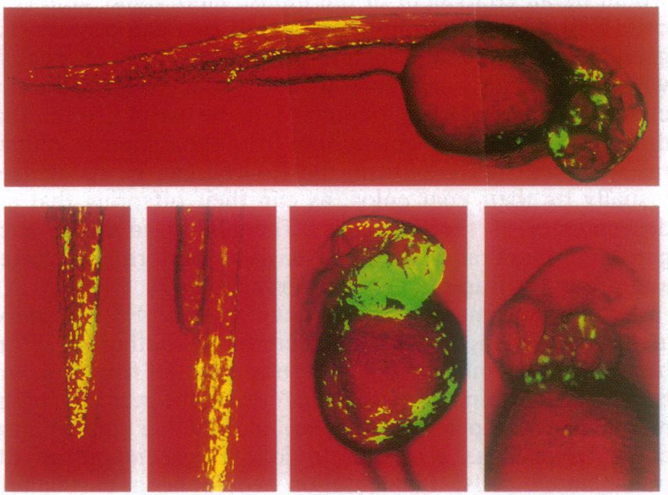
Fig. 2 Participation of transplanted cells in chimeras by 24 hr. Eggs that were to serve as donors were microinjected with fluorescein dextran. After incubation at 27°C overnight the chimeric embryos were examined and photographed separately under the Nomarski channel and fluorescein channel of a confocal microscope (Bio-Rad). The two images were merged and the fluorescent donor cells were shown as green-yellow pseudocolor. Length of the full embryo is about 1.6 mm.
Acknowledgments
This image is the copyrighted work of the attributed author or publisher, and
ZFIN has permission only to display this image to its users.
Additional permissions should be obtained from the applicable author or publisher of the image.
Full text @ Proc. Natl. Acad. Sci. USA