
|
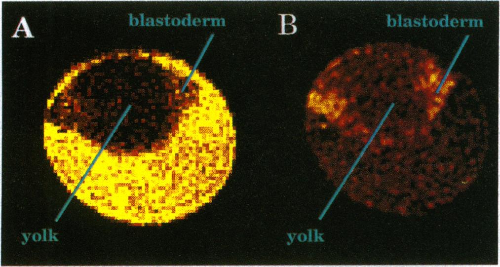
Fig. 2 False-color enhanced MR images of water in six somite dechorionated zebrafish embryos using a modified spin echo sequence (11) (128 x 128 pixels, 17 x 17 x 100 μm resolution, 3 sec repetition time; TR, the time between successive excitation pulses, two averages). Cross-sectional images were obtained through the center of embryos [diameter of six somite ca. 800 μm (6)] immersed in embryo medium held in a 1-mm glass capillary. (A) A T2-weighted image obtained using a relatively long echo time (TE, the delay between excitation and formation of the spin-echo = 55 msec) so that components with a longer T2 appear bright. This image depicted a relatively dark embryo surrounded by embryo medium (bright yellow region). The yolk appeared at the center of the embryo (dark red) with the blastoderm projecting on either side (shown in red). (B) A diffusion-weighted image obtained by applying a pair of gradient pulses (50 G/cm in magnitude, 3 msec in duration, and 3.5 msec pulse separation) around the 180° (nonslice selective) refocusing rf pulse. Spins that have diffused during the gradient pair acquire net phase shifts, resulting in a signal loss through interference. This signal loss increases with increased diffusion coefficients. These images of a zebrafish embryo reveal a relatively bright blastoderm (shown in yellow and red), dark yolk, and dark embryo medium (shown in red and black). This image indicated a lower water diffusion coefficient in the blastoderm than in the yolk and the surrounding medium. Embryos were dechorionated as previously described (12), and figures were false-color enhanced using a standard color palette from a computer graphics program (IMAGE program from National Institutes of Health).