
IMAGE
Fig. 4
- ID
- ZDB-IMAGE-080529-9
- Publication
- Slanchev et al., 2005 - Development without germ cells: The role of the germ line in zebrafish sex differentiation
- All Figures
- Figures for Slanchev et al., 2005
Image
|
Figure Caption
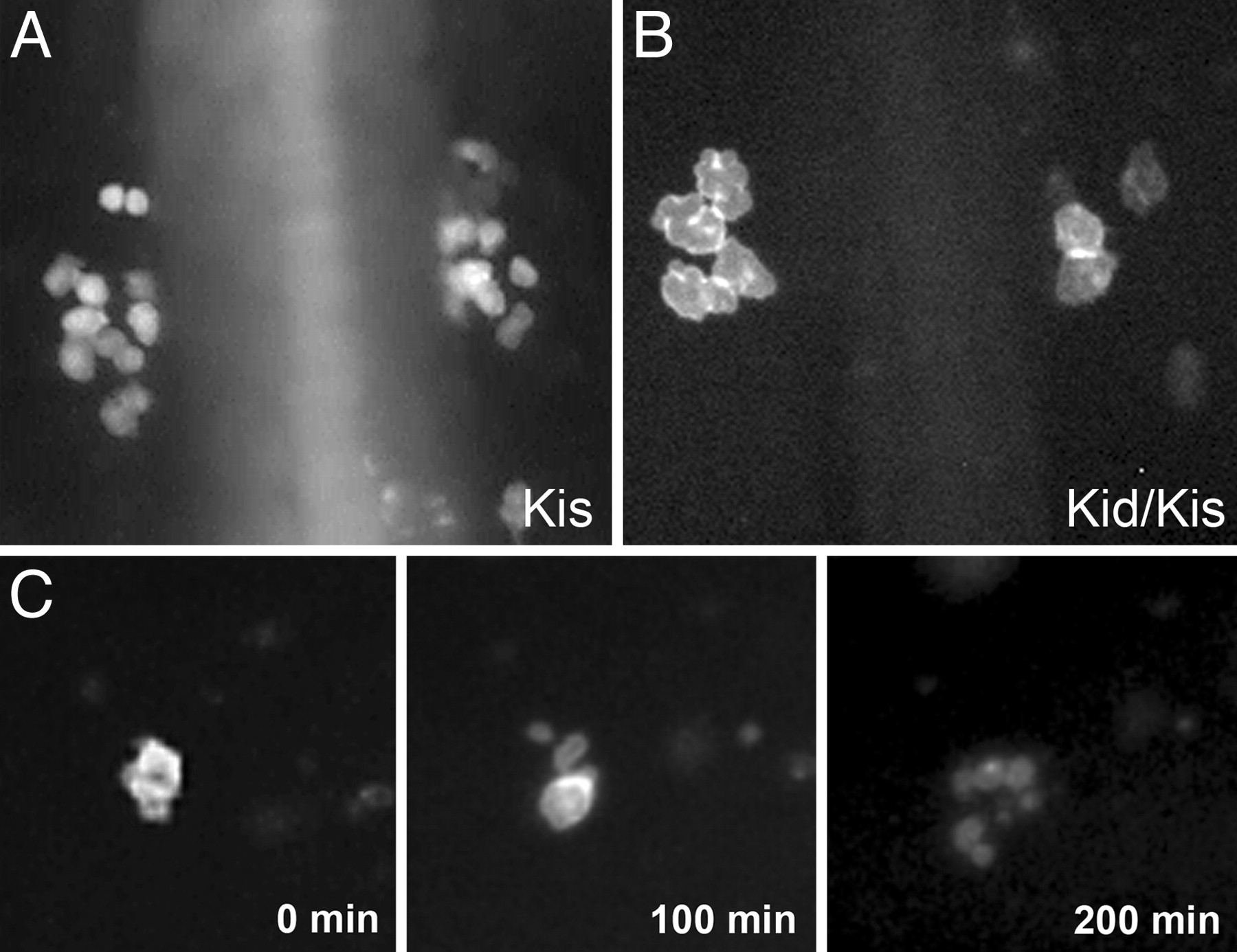
Fig. 4 PGCs in kid-nos1 3′UTR treated embryos exhibit abnormal cell morphology culminating in cell death. Fluorescently labeled germ cells in kis-glo UTR (A) and kid-nos1 3′UTR/kis-glo UTR-injected (B) embryos at 12 hpf are shown. More PGCs are found in the control embryos and these appear smaller in size relative to kid-treated PGCs. (C) Frames from a time-lapse movie of a kid-nos1 3′-UTR-treated embryo showing a PGC undergoing cell death (see Movie 2).
Acknowledgments
This image is the copyrighted work of the attributed author or publisher, and
ZFIN has permission only to display this image to its users.
Additional permissions should be obtained from the applicable author or publisher of the image.
Full text @ Proc. Natl. Acad. Sci. USA