
|
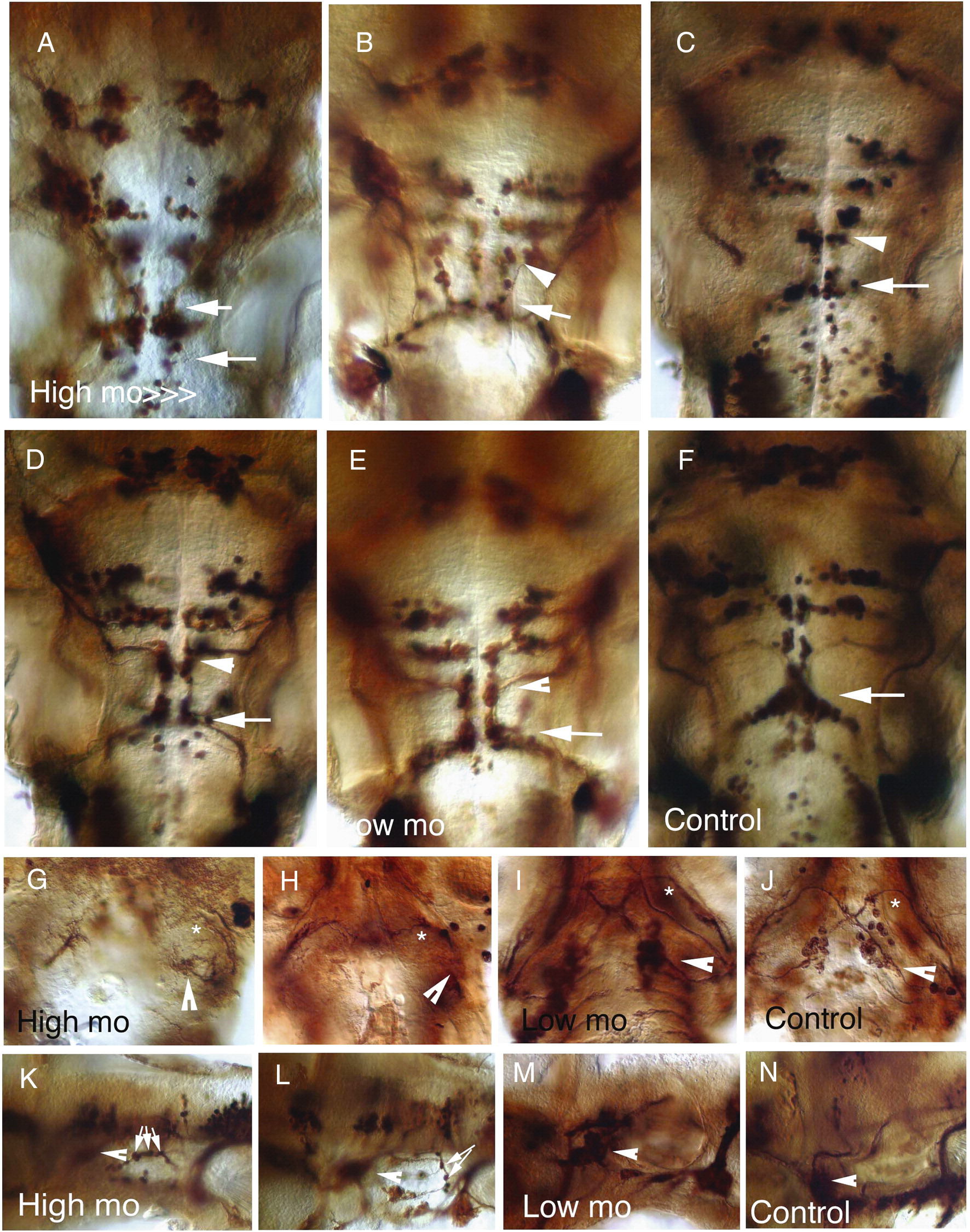
Fig. 4 Cranial motor neuron migration defects in zebrafish embryos. (A–E) 48 hpf tg(isl1:GFP) zebrafish embryos injected with decreasing amounts of tbx20 morpholino. (A) 6 ng per embryo, (B) 4 ng per embryo, (C) 3 ng per embryo, (D) 2 ng per embryo, (E) 1 ng per embryo. Dorsal views of the hindbrain, rostral is to the top. Migration defects of the nVII motor neurons (white arrowheads) that only partially reach rhombomere 6 (white arrows). (F) Uninjected control. (G–I) 5 dpf tg(isl1:GFP) zebrafish embryos injected with decreasing amount of tbx20 morpholino. Ventral views, dorsal to the top, showing the trigeminal (asterisk) and facial nerve (arrowheads). Only the facial nerves show severe pathfinding defects. (J) Uninjected control. (K–M) 5 dpf tg(isl1:GFP) zebrafish embryos injected with decreasing amount of tbx20 morpholino. Lateral views, rostral to the left. Arrowheads point to the facial sensory ganglion and adjacent facial nerve, displaying growing defects. Small white arrows point to the anterior lateral line nerve, which is not affected by tbx20 morpholino injections. (N) Uninjected control.
Reprinted from Developmental Biology, 317(2), Pocock, R., Mione, M., Hussain, S., Maxwell, S., Pontecorvi, M., Aslam, S., Gerrelli, D., Sowden, J.C., and Woollard, A., Neuronal function of Tbx20 conserved from nematodes to vertebrates, 671-685, Copyright (2008) with permission from Elsevier. Full text @ Dev. Biol.