
|
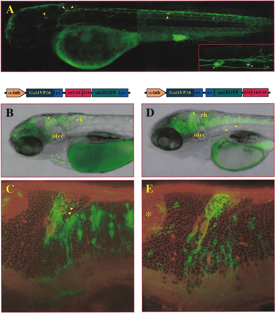
Fig. 7 Neural-specific expression in transient transgenic Gal4-VP16-activator/effector injected zebrafish embryos. Lateral views of -1696α1TIpEGFP- (A), tub-GVP-Uunc- (B, C), and tub-GVP-Uunc-inv-injected (D, E) embryos showing single optical sections which were taken at 2 (A) or 3 dpf (B–E) using confocal microscopy. Pictures represent pseudocolored composites with EGFP fluorescence displayed in green overlaid with either a transmitted light image (A, B, D) or Bodipyceramide-TexasRed counterstain to visualize overall morphology (C, E). (A) The lateral view of -1696α1TIpEGFP-injected embryo demonstrates that EGPF expression is almost exclusively confined to neurons (arrowheads). Only very few neurons show EGFP fluorescence at detectable levels. Diffusion of EGFP into the neuronal processes also visualizes axons, dendrites, and synaptic butons (see inset, magnification of EGFP-expressing neuron in trunk). (B, D) Overview of the head region of tub-GVP-Uunc- (B) and tub-GVP-Uunc-inv-injected (D) embryos, note the strong and high-frequent expression in the hindbrain and spinal chord. The tub-GVP-Uunc-inv-injected specimen (D) displays expression in the optic tectum demonstrating the linear arrangement of these neurons projecting into the tectal neuropil (*). Also, ventrally projecting axons leaving the spinal cord can be observed (e.g., arrowheads). (C, E) Higher magnification of the cerebellum (lateral views) of the injected embryos shown in (A) and (C). In the tub-GVP-Uunc-injected specimen (C), even tiny neurons (supposedly granule cells, arrowheads) and their processes can be visualized. In the tub-GVP-Uunc-inv-injected specimen (E), the predominant membrane localization of the unc76:EGFP-fusion protein can be seen highlighting the ventral projections (see arrowhead) of the labeled cerebellar neurons. Abbreviations: c, cerebellum; otec, optic tectum; rh, rhombencephalon.
Reprinted from Developmental Biology, 233(2), Köster, R.W. and Fraser, S.E., Tracing transgene expression in living zebrafish embryos, 329-346, Copyright (2001) with permission from Elsevier. Full text @ Dev. Biol.