
Image
|
Figure Caption
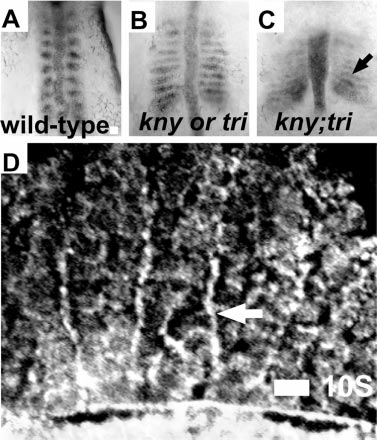
Fig. 7 Fak protein localizes to somite boundaries in kny;tri double-mutant embryos that have somites comprised of only two rows of epithelial border cells. (A–C) In situs for fak mRNA expression in 9-somite embryos (dorsal views with anterior up). (A) Wild-type embryo. (B) knypek or trilobite embryo. (C) knypek; trilobite embryos. (D) Immunostaining for Fak protein in a 10- somite kny;tri embryo (dorsal view with anterior left). Scale bars, 20 μm.
Acknowledgments
This image is the copyrighted work of the attributed author or publisher, and
ZFIN has permission only to display this image to its users.
Additional permissions should be obtained from the applicable author or publisher of the image.
Reprinted from Developmental Biology, 240(2), Henry, C.A., Crawford, B.D., Yan, Y.-L., Postlethwait, J., Cooper, M.S., and Hille, M.B., Roles for zebrafish focal adhesion kinase in notochord and somite morphogenesis, 474-487, Copyright (2001) with permission from Elsevier. Full text @ Dev. Biol.