
|
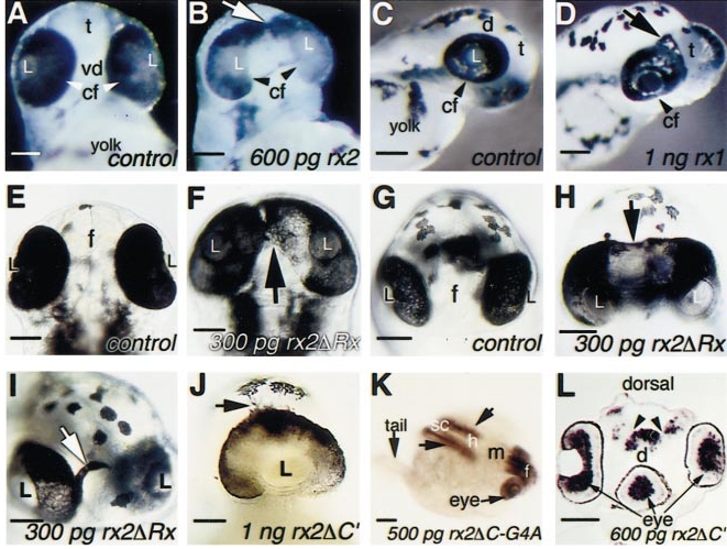
Fig. 2 Eye phenotypes and axis duplication in embryos injected with rx1 or rx2 RNA. (A-J) Comparison of wild type (A, C, E, G) and rx1- or rx2-injected embryos (B, D, F, H–J) at 60 h postfertilization (hpf), in a ventral view (A, B, E, F, J), a lateral view (C, D), or a frontal view (G-I). Arrows indicate the medial expansion of retinal tissue toward forebrain territories. (K) Partial axis duplication (arrows) caudal to the midbrain. (L) Transverse section showing a third eye in the ventral midline (arrow) and a partially duplicated forebrain (arrowheads). (K) and (L) were hybridized with a pax6.1 RNA probe and viewed in a whole-mount (K, 60 hpf) or in a cryosection (L, 26 hpf). cf, choroid fissure; d, diencephalon; f, forebrain; h, hindbrain; L, lens; m, midbrain; sc, spinal cord; t, telencephalon; vd, ventral diencephalon. Scale bars: 100 μm.
Reprinted from Developmental Biology, 231, Chuang, J.-C. and Raymond, P.A., Zebrafish genes rx1 and rx2 help define the region of forebrain that gives rise to retina, 13-30, Copyright (2001) with permission from Elsevier. Full text @ Dev. Biol.