
|
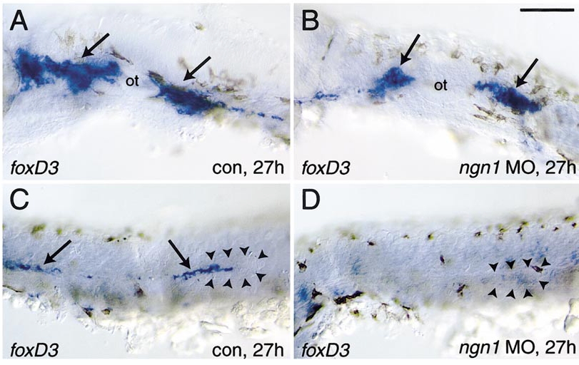
Fig. 8 Glial cell differentiation after ngn1 morpholino injection. (A, B) Neural crest-derived glial cells initially differentiate despite the lack of cranial ganglia. In control embryos (A), glial cells found in association with cranial ganglia (arrows) on either side of the otic vesicle (ot) express foxd3. In injected embryos, glial cells are found in approximately the same positions, although sometimes foxd3 expression is lower. (C) Glial cells (arrows) are closely associated with the lateral line nerve and migrating primordium (outlined by arrowheads), but are missing after ngn1 morpholino injection. Bar, 100 μm.
Reprinted from Developmental Biology, 251(1), Andermann, P., Ungos, J., and Raible, D.W., Neurogenin1 defines zebrafish cranial sensory Ganglia precursors, 45-58, Copyright (2002) with permission from Elsevier. Full text @ Dev. Biol.