
|
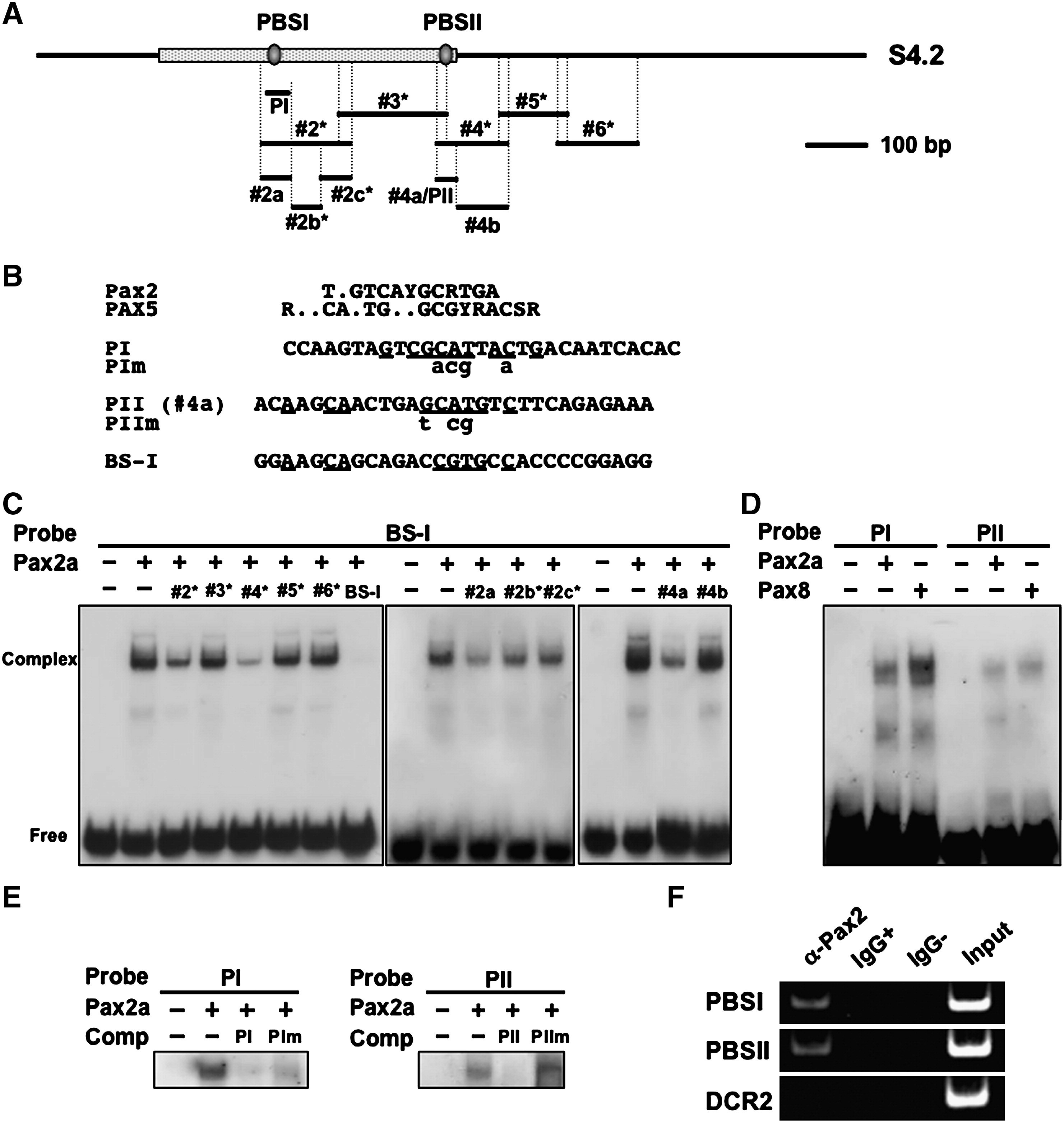
Fig. 7 Binding of Pax2a to the S4.2 enhancer in vitro and in vivo. (A) Subfragments of the S4.2 region examined using EMSA in panels C–E. See Fig. 5A for sequences #2a, #2b*, and #2c*. (B) Alignment of the sequences of the oligos for PBSI (PI), PBSII (PII), and BS-I (Song et al., 1996) with the consensus sequences for Pax2 (Epstein et al., 1994) and Pax5 (Czerny et al., 1993). Conserved nucleotides are underlined. Base substitutions in the mutated forms of PI and PII (PIm and PIIm) are indicated in lowercase. (C) Binding of Pax2a with the reference probe, BS-I, was challenged using a 100-fold molar excess of the respective subfragments as competitors. (D) Direct binding of PBSI and PBSII to Pax2a and Pax8 using DIG-labeled PI and PII probes. (E) Binding of Pax2a to DIG-labeled PI and PII was competed efficiently by a 100-fold molar excess of the corresponding DNA, but their competing activity was abrogated by base substitutions within the core binding sequences (PIm and PIIm). (F) The regions covering PBSI and PBSII were amplified separately from immunoprecipitates obtained by treating the sonicated chromatin of 16-somite-stage embryos with anti-Pax2 (ChIP assay); no amplification occurred when the chromatin was incubated with or without IgG.
Reprinted from Developmental Biology, 316(2), Inoue, F., Parvin, M.S., and Yamasu, K., Transcription of fgf8 is regulated by activating and repressive cis-elements at the midbrain-hindbrain boundary in zebrafish embryos, 471-486, Copyright (2008) with permission from Elsevier. Full text @ Dev. Biol.