
|
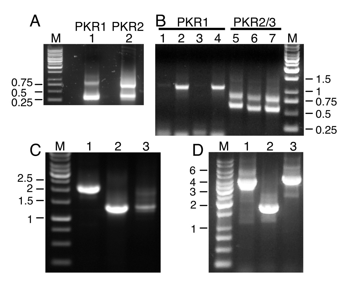
Fig. 1 PCR for cloning of T. nigroviridis PKR genes. (A) Results for 5′ RACE PCRs with T. nigroviridis cDNA are shown for primers specific for PKR1 (lane 1) and PKR2 (lane 2). M denotes the 1 kb marker. Fragments are labeled in kb. (B) shows the results of 3′ RACE PCR using four different forward primers used in primary and nested PCRs for PKR1 (lanes 1–4) and three different forward primers combinations for PKR2 and PKR3 (lanes 5–7). The smaller fragment represents PKR2 and the larger one represents PKR3. (C) PCR products are shown using primers covering the complete open reading frames of PKR1 (lane 1), PKR2 (lane 2) and PKR3 (lane 3). (D) PCR reactions of overlapping regions were performed with genomic DNA to elucidate the genomic organization of PKR1. Lanes 1 and 2 show the PCR products obtained with primers spanning the region between the 5′ untranslated region of PKR1 and exon 15 and exon 14 and the 3′ untranslated region of exon 19, respectively. PCR product shown in lane 3 was obtained with primers covering exon 17 of PKR1 and exon 7 of PKR2. PCR products were cloned and completely sequenced.