
Image
|
Figure Caption
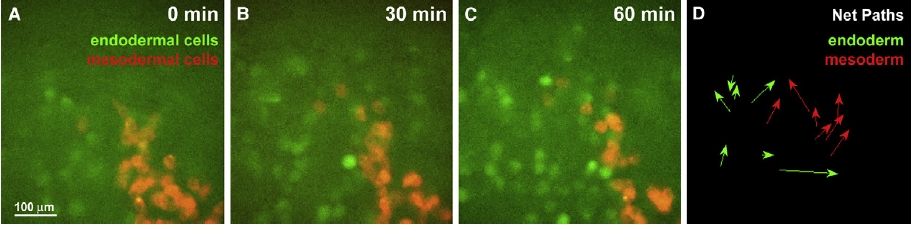
Fig. S2 Endodermal Cells Do Not Behave Like Mesodermal Cells during Early Gastrulation (A–C) Shown is the evolution of a clone of mesodermal cells (labeled in red) transplanted at the margin of an Et(CLG-YFP)smb602 embryo (in green) from 60% to 70% epiboly (lateral view, animal pole to the top, dorsal to the right). (D) Net path of internalized mesodermal cells (red) and endodermal cells (green) from time lapse in (A)–(C) is shown. Whereas mesodermal cells move toward the animal pole, endodermal cells do not show any preferred direction.
Acknowledgments
This image is the copyrighted work of the attributed author or publisher, and
ZFIN has permission only to display this image to its users.
Additional permissions should be obtained from the applicable author or publisher of the image.
Full text @ Curr. Biol.