
|
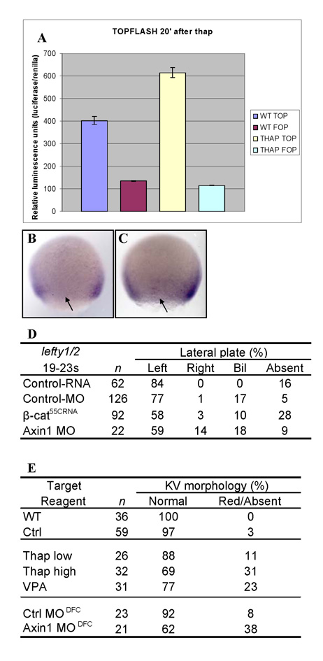
Fig. S5 Increased β-catenin reporter activity after thapsigargin treatment. (A) TopFlash (top) vs FopFlash (fop) luciferase reporter constructs analyzed for relative luminescence in wt or thap-treated embryos 20 minutes after treatment, normalized to renilla luciferase. TOPdGFP reporter transgenic line, WMISH with GFP probe, dorsal side shown, GFP expression is excluded from DFC region in (B) wt, but detected in (C) thapsigargin-treated embryos. Typical lateral domains of TOPdGFP reporter expression are present in both untreated and treated embryos. Arrows note DFC region. (D) Summary of lefty1/2 expression in DFC-targeted embryos. Axin1-MO injected had a P value <0.01 by Chi-square analysis. (E) Summary table of KV alterations of DFC-targeted MOs and Ca2+/ PI cycle inhibitors. Thap, thapsigargin; WT, wild type; Ctrl, control DMSO; Thap low, low concentration; Thap high, high concentration; VPA, valproate.