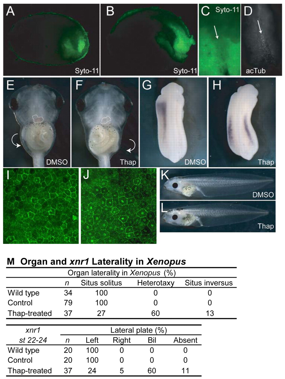
Fig. 6 Xenopus DFC-like cells and Ca2+ requirement for laterality. Xenopus embryos incubated with Syto-11 at stage 11.5 and sagittally bisected at (A) stage 14 and (B) stage 17 showing Syto-11-labeled cells enriched in the future tail region. (C) Stage 17 archenteron roof explant, showing Syto-11 staining of the node (arrow) and surrounding LPM. (D) Explant in C immunostained for acetylated tubulin (acTub), which is enriched in the node region (arrow). (E) Ventral view of stage 46 control-treated embryo, showing situs solitus, or normal lateral asymmetry. The heart is outlined, showing the left-sided position of the ventricle and right-sided outflow tract. The gut coils counterclockwise, as shown by the arrow. (F) Ventral view of a thapsigargin-treated embryo at stage 46, showing an example of situs inversus, or reversed laterality. The heart is outlined, showing the right-sided position of the ventricle and left-sided outflow tract. The gut coils clockwise, as shown by the arrow. (G) Left-sided xnr1 expression at stage 22-24 in a DMSO-treated embryo. (H) Bilateral xnr1 expression in a thapsigargin-treated embryo. (I,J) Lateral view of β-catenin protein localization in control-treated (I) and thapsigargin-treated (J) stage 13 embryos. Note increased number and intensity of nuclear staining. (K,L) Lateral views of control (K) and thapsigargin-treated (L) embryos, showing normal axial development. (M) Summary of Xenopus laterality data. Thap, thapsigargin.
Image
Figure Caption
Acknowledgments
This image is the copyrighted work of the attributed author or publisher, and
ZFIN has permission only to display this image to its users.
Additional permissions should be obtained from the applicable author or publisher of the image.
Full text @ Development

