
|
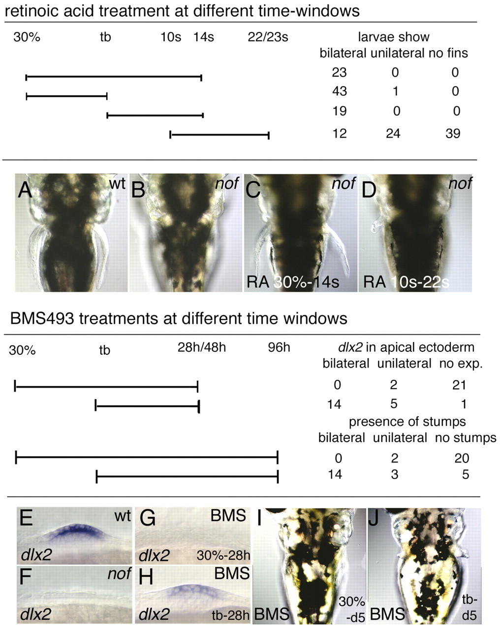
Fig. 6 (A-D) Dorsal view of the pectoral fin region on day 5 in (A) wild-type siblings and (B-D) nof mutant embryos. nof homozygotes were either (B) not treated or (C,D) treated with 10–9 M retinoic acid during the time windows indicated in the table above A-D. (E-H) Ventral view of the pectoral fin buds of (E) wild-type sibling, (F) nof and (G,H) BMS493-treated wild-type embryos. (E,H) Fin buds express dlx2, indicating AER activity in the apical ectoderm. (F,G) Fin bud regions do not express dlx2, indicating lack of AER activity in the ectoderm. (I,J) Dorsal view of the pectoral fin region of BMS493-treated wild-type embryos. Embryos were exposed to 10–6 M BMS493 at the different time windows indicated in the table above E-J.