
|
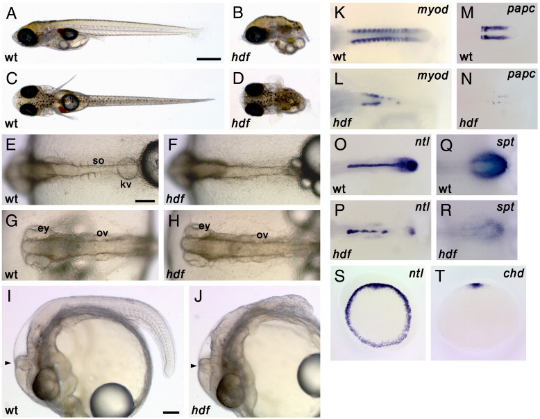
Fig. 1 Morphology and gene expression in headfish mutant embryos. (A–J) External morphology. Wild-type (A, C) and hdf mutant (B, D) embryos at hatching stage (st. 39). Lateral view (A, B) and dorsal view (C, D). Mutant embryos lack most of the posterior part of the body. On the other hand, head development appears normal. Wild-type (E, G) and hdf mutant (F, H) embryos at 4-somite stage (st. 20). Dorsal view of the tail (E, F) and head (G, H). Mutant embryos have a midline structure but fail to form somites. Lateral view of wild-type (I) and hdf mutant (J) embryos at 30-somite stage (st. 28). hdf mutant embryos show agenesis of the posterior structures but have a morphologically normal head, including the eyes, ventricles and MHB (arrowhead). ey, eye; kv, Kuppfer's vesicle; ov, otic vesicle; so, somite. Scale bars: (A) 500 μm; (E) 100 μm; (I) 100 μm. (K–T) Gene expression patterns. Wild-type (K, M, O, Q) and mutant (L, N, P, R) embryos during early segmentation stages. (K, L) The expression of myod, a somite differentiation marker, is observed in each somite in a wild-type embryo, in a bilateral regular stripe pattern (K), whereas it is severely reduced in the mutant embryos (L). (M, N) The expression of papc, a presomitic mesoderm marker, is lost and only faint expression is observed in the mutant (N). (O, P) The expression of ntl, a pan-mesodermal marker. ntl expression is detected in the notochord and tailbud in wild-type (O), but is down-regulated in tailbud and exhibits a interrupted pattern in midline of the mutant embryos (P). (Q, R) The expression of spt, a marker for the tailbud. While spt is expressed in the tailbud of the wild-type embryo (Q), it is considerably weaker in the mutant embryo (R). (S, T) Early gastrula stage. Animal-pole views are shown. ntl is expressed uniformly in the blastoderm margin of all sibling embryos obtained from a heterozygous mating (S) and chordin, a dorsal mesodermal marker is detected in the dorsal region of all sibling embryos (T). Each genotype is indicated as wt, wild-type and hdf, headfish.
Reprinted from Developmental Biology, 304(1), Yokoi, H., Shimada, A., Carl, M., Takashima, S., Kobayashi, D., Narita, T., Jindo, T., Kimura, T., Kitagawa, T., Kage, T., Sawada, A., Naruse, K., Asakawa, S., Shimizu, N., Mitani, H., Shima, A., Tsutsumi, M., Hori, H., Wittbrodt, J., Saga, Y., Ishikawa, Y., Araki, K., and Takeda, H., Mutant analyses reveal different functions of fgfr1 in medaka and zebrafish despite conserved ligand-receptor relationships, 326-337, Copyright (2007) with permission from Elsevier. Full text @ Dev. Biol.