
Image
|
Figure Caption
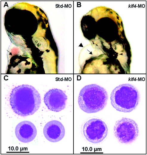
Fig. 1 Anemia and impaired erythroid cell differentiation in klf4 morphants. Anemia and pericardial oedema in klf4-MO injected embryos (B) compared with Std-MO injected controls (A). May–Grunwald–Giemsa stained blood smears show a mixture of immature (top row) and maturing (bottom row) primitive erythroid cells in Std-MO injected controls (C) but only immature erythroid cells in klf4-MO injected embryos (D).
Figure Data
Acknowledgments
This image is the copyrighted work of the attributed author or publisher, and
ZFIN has permission only to display this image to its users.
Additional permissions should be obtained from the applicable author or publisher of the image.
Reprinted from Mechanisms of Development, 124(9-10), Gardiner, M.R., Gongora, M.M., Grimmond, S.M., and Perkins, A.C., A global role for zebrafish klf4 in embryonic erythropoiesis, 762-774, Copyright (2007) with permission from Elsevier. Full text @ Mech. Dev.