
|
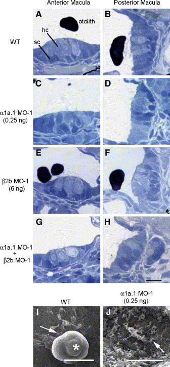
Fig. 9 Histological analysis of α1a.1 and β2b morphant ears. (A–H) 72 hpf wild type (WT) and morphant embryos were fixed, embedded in resin, sectioned at 1 μm, and stained with Toluidine blue. (A, C, E, and G) Sections through the anterior macula. (B, D, F, and H) Sections through posterior macula. Embryos injected with 0.25 ng of α1a.1 MO-1 are shown in panels C and D. Embryos injected with 6 ng of β2b MO-1 are shown in panels E and F, while embryos coinjected with 0.125 ng of α1a.1 MO-1 and 2 ng of β2b MO-1 are shown in panels G and H. hc, hair cell; sc, supporting cell. (I and J) Scanning electron micrographs of posterior macular otolithic membrane of WT embryo (I) and α1a.1 morphant (J) (injected with 0.25 ng α1a.1 MO-1) at 72 hpf. Arrows indicate hair cell ciliary bundles. Asterisk indicates otolith. Scale bars: 10 μm.
Reprinted from Developmental Biology, 294(1), Blasiole, B., Canfield, V.A., Vollrath, M.A., Huss, D., Mohideen, M.A., Dickman, J.D., Cheng, K.C., Fekete, D.M., and Levenson, R., Separate Na,K-ATPase genes are required for otolith formation and semicircular canal development in zebrafish, 148-160, Copyright (2006) with permission from Elsevier. Full text @ Dev. Biol.