
Image
|
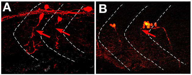
Figure Caption
Fig. S2 Abnormal axonal pathfinding of putative motoneurons in the spinal cord of vmc Isl1-GFP embryos at 36 hpf. (A,B) Dorsal, top; anterior, left. The axons of these neurons were visualised by stochastic expression of Kaede using the HuC-Kaede construct with direct fluorescent microscopy. Their axons grew anteriorly within the spinal cord, then extended out of the spinal cord through abnormal exit points (arrows).
Figure Data
Acknowledgments
This image is the copyrighted work of the attributed author or publisher, and
ZFIN has permission only to display this image to its users.
Additional permissions should be obtained from the applicable author or publisher of the image.
Full text @ Development