
|
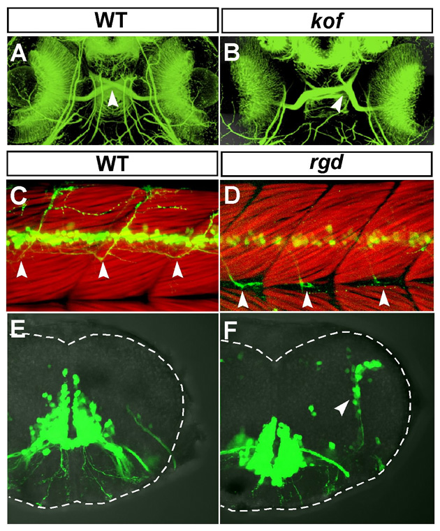
Fig. S1 Additional defects in the kof and rgd mutant embryos in the developing central nervous system. (A,B) Ventral views of the head region of 72-hpf wild-type (A) and kof (B) embryos. Anterior, top. The retinal axons were stained with the anti-acetylated α-tubulin antibody. In the wild-type embryos, the optic chiasm was formed in the ventral midline (A, arrowhead). In the kof embryos, the position of the optic chiasm was randomly shifted toward the left or right near the midline (B, arrowhead). (C,D) Lateral views of the trunk region of 72-hpf wild-type (C) and the rgd (D) embryos. Dorsal, top; anterior, left. The trunk muscles were visualised with rhodamine-phalloidin. In the wild-type embryos, Isl1-GFP-positive spinal motor axons extended to the dorsal part of the trunk muscles (C, arrowheads). In the rgd embryos, Isl1-GFP-positive spinal motor axons grew ventrally and changed their growth pathway posteriorly after reaching the level of the myoseptum (D, arrowheads). (E,F) Transverse views of the hindbrain at r6 of the 72-hpf wild-type (E) and rgd (F) embryos. Dorsal, top. In the wild-type embryos, there were no Isl1-GFP-positive cells in the lateral part of the hindbrain (E). In the rgd embryos, Isl1-GFP-positive cells were ectopically observed in the lateral part of the hindbrain (F, arrowhead).