
Image
|
Figure Caption
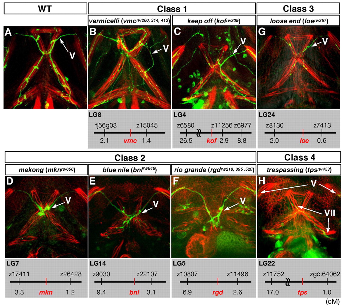
Fig. 3 The projection patterns to the ventral jaw muscles of mutants displaying defects in the outgrowth of the Vp and VII motor axons. Ventral views of the lower jaw of 72-hpf wild-type (A) and the four classes of mutant (B-H) zebrafish embryos. Anterior, top. The cranial motor axons were labelled with the Isl1-GFP transgene, and each jaw muscle was stained with rhodamine-phalloidin. The genetic locus of each mutation is shown below each panel.
Acknowledgments
This image is the copyrighted work of the attributed author or publisher, and
ZFIN has permission only to display this image to its users.
Additional permissions should be obtained from the applicable author or publisher of the image.
Full text @ Development