
|
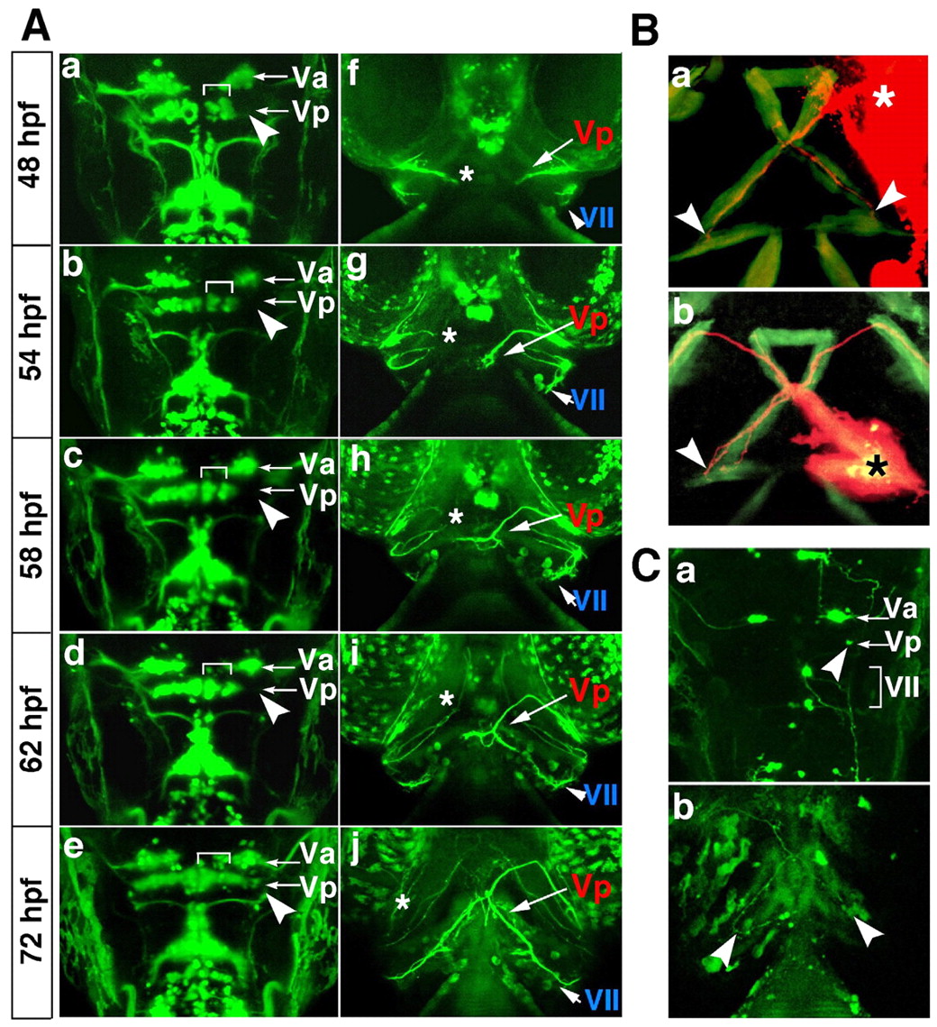
Fig. 2 No interaction is required between the left and right Vp motor axons for projection to the bilateral interhyal muscles in BA2. (A) Time-lapse images from 48-72 hpf of the Vp motoneurons after laser irradiation to the lateral part of the Vp motoneurons on the right side (a-e, arrowheads). Dorsal (a-e) and ventral (f-j) views of the same operated zebrafish embryo. Anterior, top. Brackets indicate remaining cells on the right side (a-e). Asterisks indicate the nerve end of the operated motoneurons (f-j). (B) Anterograde (a) and retrograde (b) labelling of the Vp motor axons of 72-hpf wild-type embryos with DiI. Ventral view; anterior, top. Asterisks indicate the points of DiI application. Arrowheads indicate the ends of the labelled axons. (C) Stochastic expression of GFP using the Isl1-GFP construct. Ventral (a) and dorsal (b) views; anterior, top. Arrowheads indicate a single Vp motoneuron labelled with the Isl1-GFP construct (a) and the ends of the labelled axons (b).