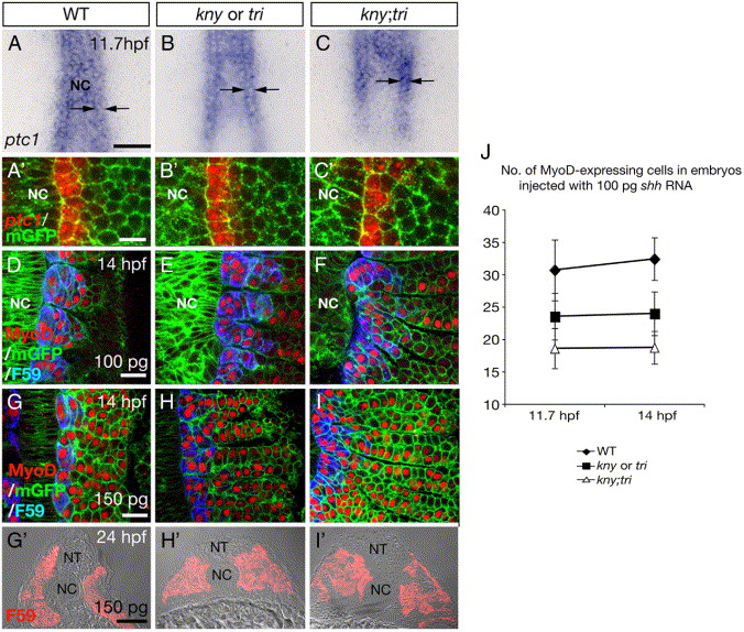
Fig. 5 The range of Hh signaling in the PSM declines during early segmentation. (A–C, A′–C′) ptc1 RNA expression was restricted to one column of cells next to the notochord at the 5-somite stage in WT (A, A′), individual (B, B′), and kny;tri double mutants (C, C′). Arrows show the ML range of the ptc1 expression. (D–F) Labeling of MyoD and F59 antibodies in embryos injected with 100 pg synthetic shh RNA to induce ectopic Shh activity. (G–I) In WT, individual mutants and double mutants, injection of 150 pg shh RNA caused strong MyoD protein expression throughout the somite at the 10-somite stage. (G′–I′) Transverse sections of day1-embryos injected with 150 pg shh RNA and labeled with F59 antibody. (J) Numbers of MyoD-expressing cells at the 5-somite stage and numbers of cells co-labeled by MyoD and F59 antibodies at the 10-somite stage in embryos injected with 100 pg shh RNA. For each genotype, 10 embryos were examined at both stages and quantifications were all done within the 3rd somite. Error bars represent the standard deviation. (A–I) Dorsal views. NC, notochord. Scale bars: (A–C, G′–I′) 100 μm, (A′–C′) 20 μm, (D–I) 50 μm.
Reprinted from Developmental Biology, 304(1), Yin, C., and Solnica-Krezel, L., Convergence and extension movements mediate the specification and fate maintenance of zebrafish slow muscle precursors, 141-155, Copyright (2007) with permission from Elsevier. Full text @ Dev. Biol.

