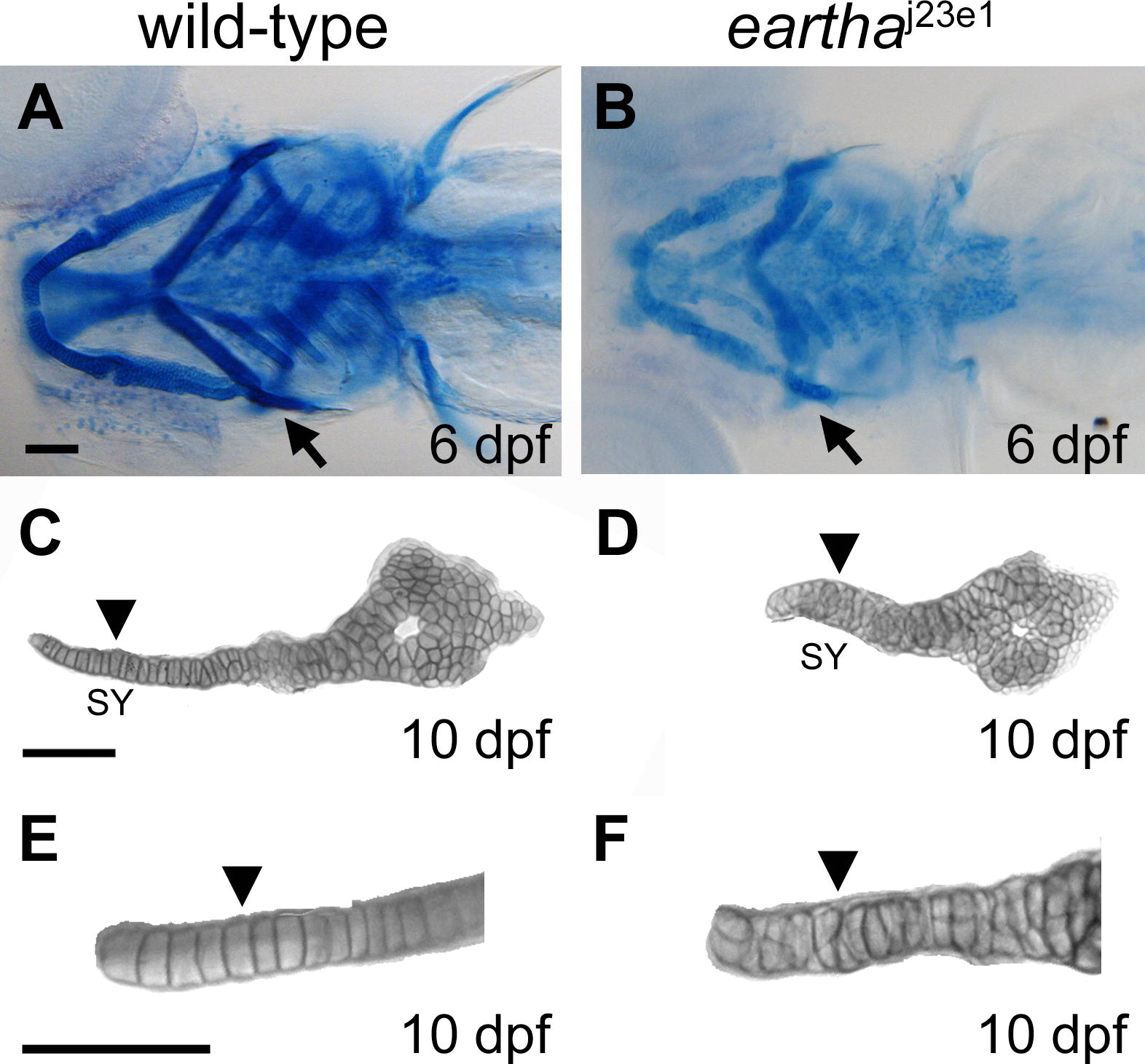
Fig. 3 The earthaj23e1 Mutation Affects Cartilage Maturation (A–F) The pharyngeal skeleton of wild-type and earthaj23e1 larvae are revealed by Alcian Blue staining, where the staining in earthaj23e1 larvae (B) is significantly weaker than that in wild-type larvae (A). At 10 dpf, the chondrocytes in the symplectic (SY) region (arrowhead in [C] and [E]) of the hyosymplectic cartilage are arranged into a single cell wide stack (arrowhead in [E]) in wild-type larvae. This organization of chondrocytes is disarranged in the earthaj23e1 larvae (arrowheads in [D] and [F]). Arrows in (A) and (B) indicate the hyosymplectic cartilages that are shown in (C) and (D). Arrowheads in (C) and (D) mark the SY region of hyosymplectic cartilage. (E) and (F) are enlarged images of the SY region of hyosymplectic cartilage. Scale bars: 100 μm.
Image
Figure Caption
Figure Data
Acknowledgments
This image is the copyrighted work of the attributed author or publisher, and
ZFIN has permission only to display this image to its users.
Additional permissions should be obtained from the applicable author or publisher of the image.
Full text @ PLoS Genet.

