
|
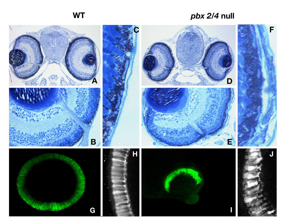
Fig. 1 The laminar structure of pbx2/4 null eyes is normal after 5 days of development. All laminar layers are present and in both wild type (A and B) and, and lzr mutants injected with pbx2/4 morpholinos (D and E). We note a consistent decrease in eye size in pbx2/4 null embryos, but the proportion of eye area occupied by the retinal pigmented epithelium in morphants is significantly increased (F verses C). The relative position of the optic nerve is similar in both wild type and morphant embryos. The retinal photoreceptor layer is absent in the ventral domain of the eye in lzr mutants injected with pbx2/4 morpholinos (I) when compared to wildtype (G) and is disorganized (compare H, wildtype, with J, lzr mutants injected with pbx2/4 morpholinos). Photoreceptor labeling was accomplished using the zpr-1 monoclonal antibody.