
|
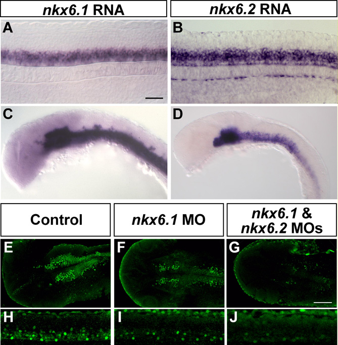
Fig. S2 Anti-Nkx6 detects both Nkx6.1 and Nkx6.2 proteins. 24-hpf embryos, anterior to the left. (A-D,H-J) lateral views; (E-G) dorsal views. The confocal images in E-G were all taken at the same settings to ensure comparability; likewise for the confocal images in H-J. The confocal images in H-J show only one side of the spinal cord; thus, there are fewer labeled cells than in the images in A and B, which show whole-mount embryos. nkx6.1 (A) and nkx6.2 (B) mRNA are expressed in the ventral spinal cord in similar patterns. Both nkx6.1 and nkx6.2 (C,D) mRNA are expressed in the ventral brain, although the expression patterns are different. Injection of nkx6.1 MO decreased Nkx6.1 antibody labeling in brain (F) and spinal cord (I) as compared to controls (E,H). However, some Nkx6.1-positive cells remained, suggesting that the antibody recognizes both Nkx6.1 and Nkx6.2 proteins. We tested this hypothesis by co-injecting nkx6.1 and nkx6.2 MOs, which extinguished nearly all Nkx6 antibody labeling in the spinal cord (J) and brain (G). Thus, we infer that the antibody recognizes both Nkx6.1 and Nkx6.2 proteins. Scale bars: 50 μm.