
|
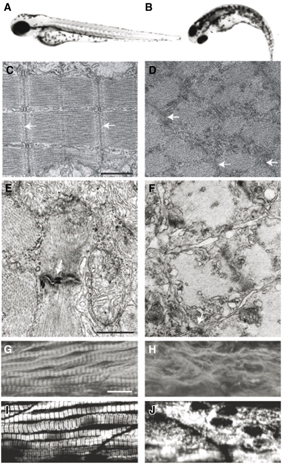
Fig. 1 Zebrafish embryos with a mutation in the steif gene fail to form myofibrils in the skeletal musculature and the heart. (A, B) 3-day-old steif mutants (B) are immotile and have smaller eyes than wild-type embryos (A). (C, D) electron microscopic analysis of parasagittal sections through a somitic fast muscle cell of a wild-type and a steif mutant embryo (48 hpf). In contrast to wild-type (C), steif mutant (D) skeletal fast muscle cells do not form ordered myofibrils. (E, F) electron micrographs of sections through cardiac muscle cells from 5-day-old wild-type (E) and mutant embryos (F). The mutant lacks myofibrils and does also not form proper intercalated disks (arrows indicate Z-lines of sarcomeres). (G, H) superficial wild-type and mutant slow muscle cells stained immunohistochemically with the antibody F59 directed against slow muscle myosin. Wild-type (G) but not the steif mutant cells (H) show the striped pattern of slow muscle myosin staining. (I, J) F-actin staining of wild-type (I) and steif (J) slow muscle cells. As in the case of the myosin-containing thick filaments, the thin filaments composed of F-actin are strongly disorganized in the mutant (J). Scale bars: 1 μm (C, D); 0,5 μm (E, F); 25 μm (G–J).
Reprinted from Developmental Biology, 308(1), Etard, C., Behra, M., Fischer, N., Hutcheson, D., Geisler, R., and Strähle, U., The UCS factor Steif/Unc-45b interacts with the heat shock protein Hsp90a during myofibrillogenesis, 133-143, Copyright (2007) with permission from Elsevier. Full text @ Dev. Biol.