
|
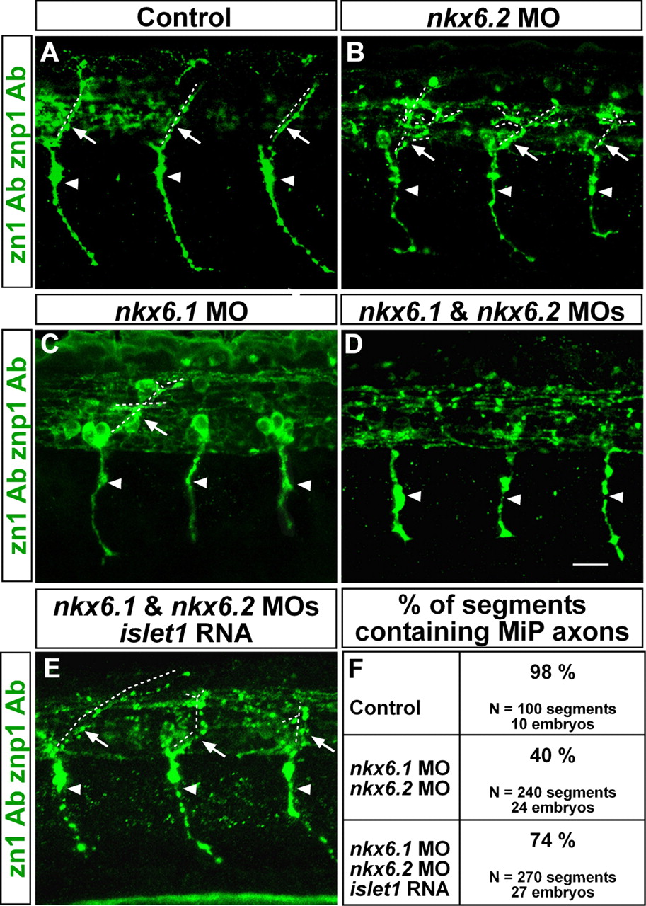
Fig. 3 MiP axons are absent from embryos lacking Nkx6 proteins. (A-D) Lateral views at axial level 8-12 of 28-hpf embryos labeled with zn1 and znp1 Abs. Arrowheads point to CaP axons; arrows point to MiP axons; dashed lines indicate MiP axon trajectories. Embryos lacking Nkx6.2 or Nkx6.1 (B,C) have normal CaP axons, but MiP axons were absent, truncated or excessively branched compared with control embryos (A). (D) Loss of MiP axons is most severe in double nkx6 MO-injected embryos, but CaP axons are normal. (E) Loss of MiP axons in embryos lacking Nkx6.1 and Nkx6.2 is restored by co-injection of islet1 mRNA with the nkx6 MOs. (F) Quantification of segments lacking MiP axons in control, nkx6 MO-injected, and nkx6 MO plus islet1 mRNA-injected embryos. Scale bar: 20 µm.