
|
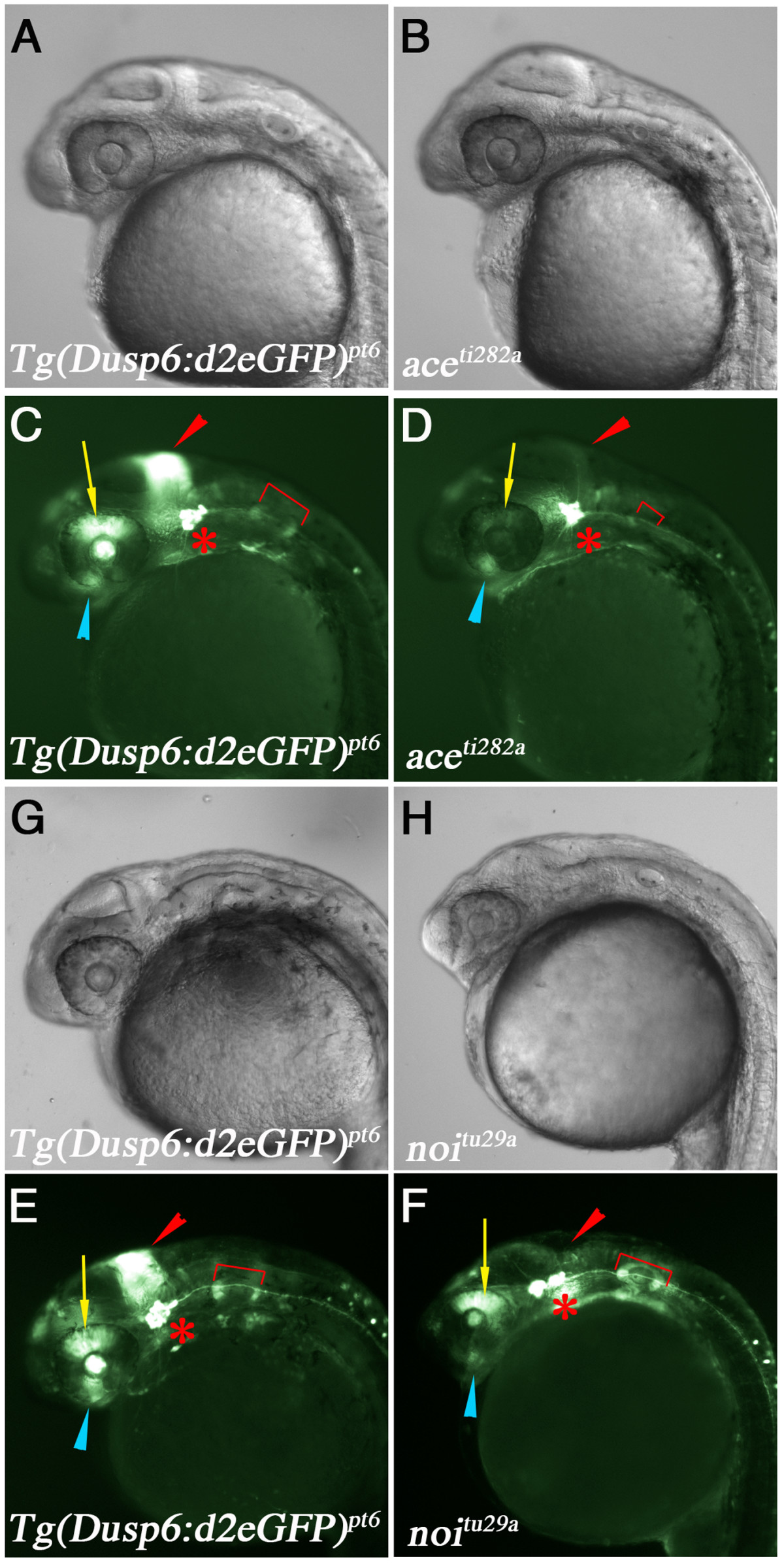
Fig. 4 D2EGFP expression in fgf8/ace and pax2.1/noi mutants. (A-F) Lateral views of Tg(dusp6:d2EGFP)pt6 embryos at 28 hpf crossed into ace or noi mutants. Genotype is listed in bottom left corner. (A & B) Brightfield images of wildtype sibling and ace mutant embryo, respectively. (C & D) d2EGFP expression in WT sibling and ace mutant. Note loss of d2EGFP expression within the MHB, dorsal retina, and the smaller otic vesicle, while d2EGFP expression in the trigeminal ganglia is unaffected. (G & H) Brightfield images of wildtype sibling and noi mutant embryo. (E & F) d2EGFP expression is lost in the MHB and optic stalk. In contrast to the ace mutants, expression within the dorsal retina and otic vesicles are normal.