
|
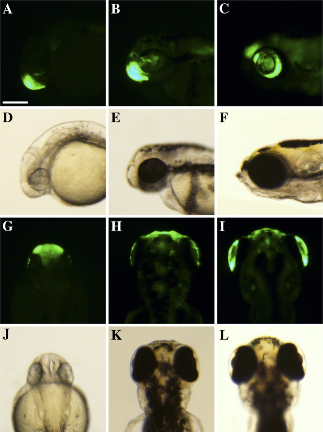
Fig. 3 Green fluorescent protein (GFP) expression of the gsnl1 6.4-kb reporter in stable transgenic embryos. A-F: Lateral view of GFP (A-C) and brightfield images (D-F). G-L: Dorsal view of GFP (G-I) and bright field images (J-L). A,D,G,J: The 1 dpf embryos showed GFP expression in the nose region, which was primarily localized at the nasal pit and in a subpopulation of the head mesenchyme cells at the most anterior part. B,E,H,K: On 3 dpf embryos, the strong GFP signal was detected in the nose and the weak signal was observed on the future cornea in a mosaic pattern. C,F,I,L: The GFP signal was detected in the annular ligament of the eye and in the corneal epithelium in patches on 5-dpf embryos. Scale bar = 200 μm.