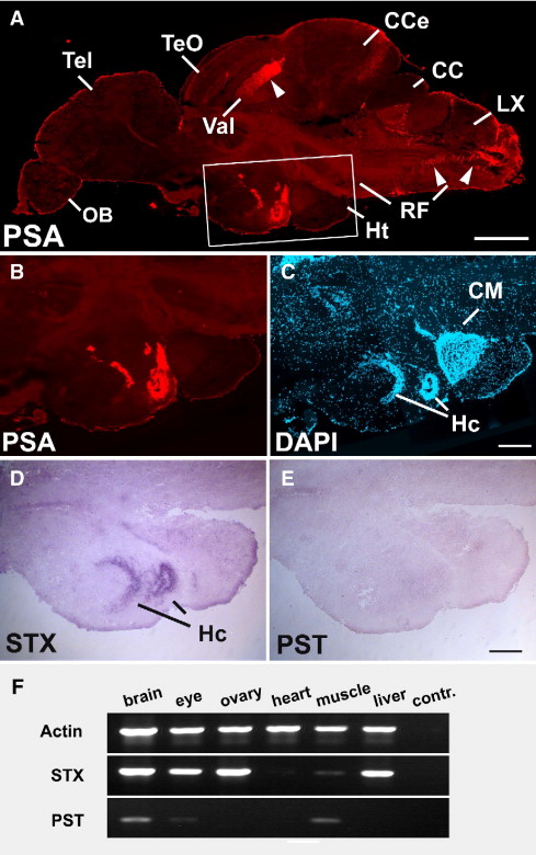
Fig. 3
Expression of PSA and zebrafish polysialyltransferase mRNAs in the adult brain. (A) Sagittal section of an adult brain, stained for PSA. Boxed region in A is enlarged in B–E. (B–E) PSA is expressed in periventricular regions of the hypothalamus (B) on cell bodies (C, nuclear stain). Only Stx is found in the Hc (D) whereas Pst is not detectable (E). (F) RT-PCR analysis on adult tissue cDNAs reveals expression of Stx in all tissues except for heart and skeletal muscle. Pst expression is evident in brain and skeletal muscle. Target amplifications were performed as described in Fig. 2. cc, crista cerebellaris; Cce, corpus cerebelli; CM, corpus mamillare; Hc, caudal zone of periventricular hypothalamus; Ht, hypothalamus; LX, vagal lobe; OB, bulbus olfactorius; RF, reticular formation; Tel, telencephalon; TeO, tectum opticum; Val, valvula cerebelli. Scale bars 500 µm (A); 200 μm (B-E).
Reprinted from Developmental Biology, 306(2), Marx, M., Rivera-Milla, E., Stummeyer, K., Gerardy-Schahn, R., and Bastmeyer, M., Divergent evolution of the vertebrate polysialyltransferase Stx and Pst genes revealed by fish-to-mammal comparison, 560-571, Copyright (2007) with permission from Elsevier. Full text @ Dev. Biol.

