
|
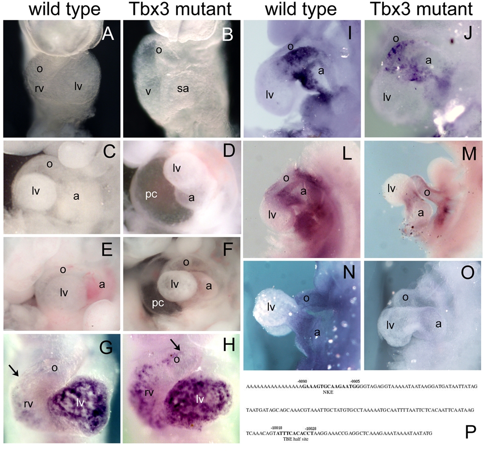
Fig. 5 (A, B) Severely affected Tbx3 mutant embryos (B) present a delay in heart looping at E9.5, in which the ventricle is still at the same dorsoventral position as the atrium. (C, D) At E10.5, the most affected Tbx3 mutant embryos display obvious heart defects, including lack of the constriction in the AVC and absence of looping in a swollen pericardiac cavity (D), compared to wild type (C). (E, F) E11.5 Tbx3 null homozygous embryos present a significant delay in heart looping and pericardiac swelling compared to wild type. (G–O) Whole mount in situ hybridization analysis at E9.5; ventral views (G, H) and lateral views (I–O). (G, H) Upon looping initiation, Bmp10 starts to be expressed in the chamber myocardium. However, Bmp10 was ectopically expressed in the non-chamber myocardium of the heart of Tbx3 mutant embryos (arrow in G, H). (I, J) Bmp2 is expressed in the AVC myocardium at E9.5 and was not altered in Tbx3 mutant embryos. (L, M) TGFβ2 is normally expressed in the non-chamber myocardium of the looping heart. In Tbx3 mutant embryos, expression of TGFβ2 is downregulated in the heart. (N, O) Smad6 is expressed in the endocardium at the level of the OFT and AVC at E9,5. However, in Tbx3 mutant embryos, expression of Smad6 is absent. (P) Consecutive NKE and TBE binding sites are found in the human BMP10 promoter, between 9800 and 10100 bp upstream the ATG codon. a, atrium; lv, left ventricle; o, outflow tract; pc, pericardiac cavity; rv, right ventricle; sa, sinoatrial region.