
|
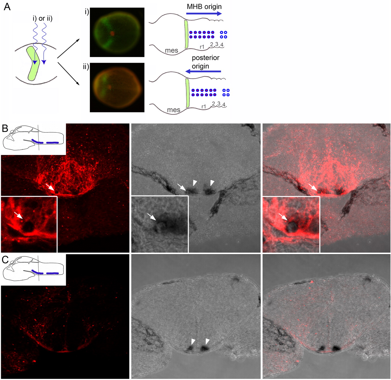
Fig. 6 Origin of rhombomere (r) 1-2 serotonergic precursors. A: Strategy for tracing the origin of r1-2 serotonergic precursors in her5PAC:egfp transgenic embryos injected with caged-fluorescein at one-cell stage (dorsal views, anterior left). An ultraviolet-light beam was focused along the midline (i) within or (ii) posterior to the green fluorescent protein (GFP) -expressing area at 90% epiboly/tail bud (left drawing; green GFP-positive midbrain-hindbrain boundary [MHB] progenitor pool). Photomicrographs: control embryos fixed directly after uncaging and processed for GFP (green) and fluorescein (red) immunostaining. Schematic pictures: two possible outcomes at 36 hours postfertilization (hpf). Top: uncaging within the GFP-positive area and pet1-positive cells of r1-2 fluorescein-labeled (red); origin within the MHB pool; Bottom: uncaging posterior to the GFP-positive area and pet1-positive cells of r1-2 fluorescein-labeled; origin posterior to the MHB pool. B: A 16-μm optical projection of a coronal cryosection through r1-2 of embryos uncaged within the GFP-expressing domain. Insets: High magnifications of 1-μm optical section of double-labeled cell indicated with an arrow. Arrowheads: pet1-positive cells on each side of the floor plate within r1-2. Note anti-fluorescein labeling of these pet1-positive cells. C: Same analysis in an embryo uncaged posterior to the GFP-expressing domain. mes, mesencephalon. Schematic picture modified from Mueller and Wullimann ([2005]).