
|
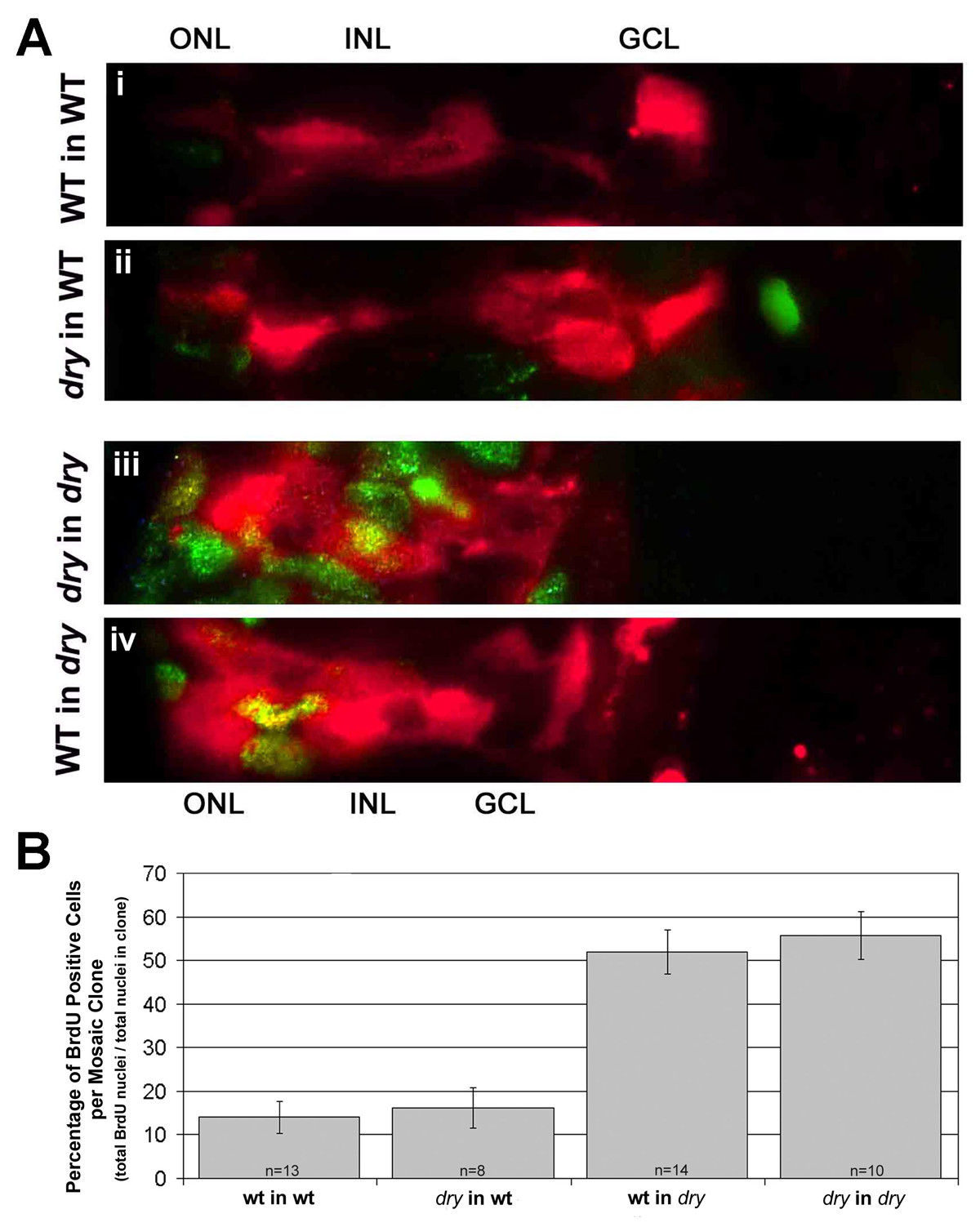
Fig. 8 Defects in cell cycle exit for disarrayed are cell-non-autonomous. (A) Genetic mosaic retinas at 61 hpf where donor cells were lineage-labeled using rhodamine conjugated dextran (red). Chimeras were injected with BrdU for one hour at 60 hpf and processed for BrdU detection (green/yellow). Donor cells are shown in the outer nuclear layer (ONL), inner nuclear layer (INL) and ganglion cell layer (GCL) regions. Cells that are double labeled with the linage marker (red) and BrdU (green) appear yellow. (i) Wild-type cells in a wild-type host embryo. (ii)disarrayed cells in a wild-type host embryo. (iii) disarrayed cells in a disarrayed mutant host embryo. (vi) Wild-type cells in a disarrayed embryo. Note the increase in proliferative cells of wild-type or disarrayed genotype in mutant retinas (iii and iv, green/yellow cells). (B) Quantization of the percent of BrdU-positive cells within small (2-20 cells) mosaic clones. n = number of mosaic clones quantitated.