
|
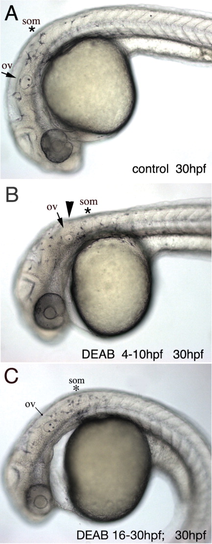
Fig. 2 Developmental defects observed in live embryos at 30 hpf when RA signaling is impaired. A: Untreated 30-hpf embryo. B: Embryos that were exposed to DEAB during gastrulation from sphere stage (4 hpf) to tailbud stage (10 hpf) show a reduction of the distance between otic vesicle (arrow) and first somite (asterisk) and a kink dorsal to the otic vesicle (arrowhead). Furthermore, the otic vesicle is smaller and only one otholith has formed. C: Embryos treated with DEAB post-gastrulation between 16-30 hpf show aberrant otic vesicle development and heart defects at 30 hpf. ov, otic vesicle; som, somites. Lateral views, anterior to the left.