
|
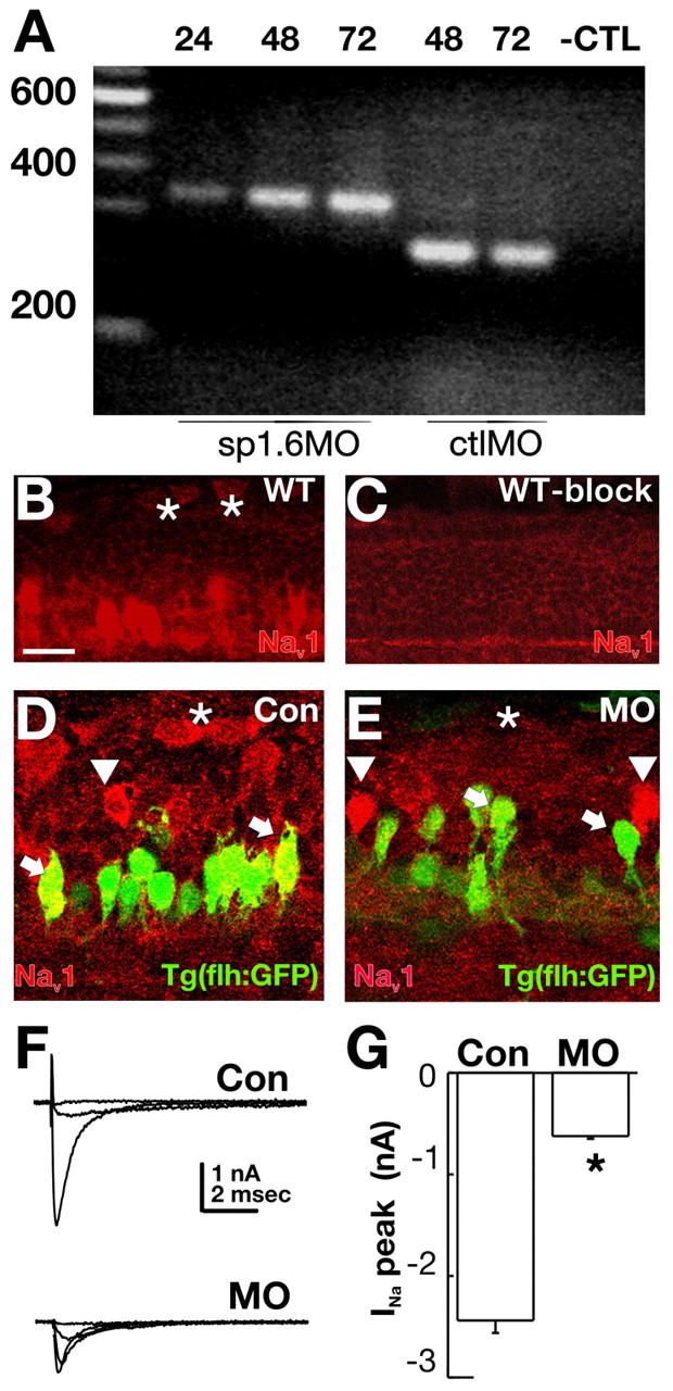
Fig. 3 Injection of 1.6MO blocked splicing and abolished Na+ channel immunoreactivity and current in spinal cord neurons that expressed scn8a mRNA. (A) In RNA isolated from embryos injected with the splice blocking MO, excision of the 96 bp intron between exons 3 and 4 did not occur. This was true at 24, 48 and 72 hpf. By contrast, embryos injected with a control splice MO displayed normal splicing at this junction. (B-E) Na+ channel immunoreactivity in control and morphant embryos. (B) A pan-specific Na+ channel antibody revealed immunoreactivity in several classes of spinal cord neurons in 48 hpf wild-type embryos. RB cells (asterisks) and ventral neurons were brightly labeled. (C) Preincubation of the antibody with peptide blocked immunodetection of Na+ channels. (D) GFP+ ventral neurons (arrowhead, arrows) in Tg(flh:GFP) embryos displayed Na+ channel immunoreactivity. (E) The 1.6MO reduced Na+ channel immunoreactivity in some (arrows, asterisks) but not all (arrowheads) spinal neurons. Scale bar: 10 μm. (F) Na+ currents recorded from RB cells of control embryos had larger amplitudes than those recorded from 1.6MO morphants. (G) Peak Na+ current amplitude was significantly reduced in RB cells of 1.6MO morphants (n=6) versus control (n=6) embryos (*P≤0.001).