
|
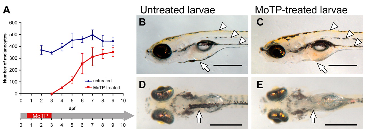
Fig. 8 Ventral yolk sac melanocytes fail to regenerate following melanocyte ablation by MoTP treatment. (A) Larval melanocytes were counted in the untreated larvae (blue line) and MoTP-treated (14-72 hpf) larvae (red line). After the appearance of melanocytes at 24 hours post-MoTP treatment, the number of melanocytes continues to steadily rise in the ensuing 4 days and then plateaus at approximately 350-400 melanocytes by 9 dpf (6 days after removal of MoTP). (B-E) At this stage of melanocyte regeneration, MoTP-treated larvae (C,E) have regenerated almost identical pigment patterns to those of the untreated larvae (B,D), with a similar number and distribution of melanocytes in the dorsal, lateral and ventral larval melanocyte stripes (white arrowheads). However, MoTP-treated larvae fail to regenerate the majority of the ventral yolk sac melanocytes (white arrow in C and E). Scale bars: 500 μm.